Ascariasis-related surgical complications still occur in India. Ascariasis usually manifests as diarrhoea, abdominal pain, and anaemia. Bowel obstruction is a frequent cause of emergency presentations related to ascariasis in developing countries. However, bowel perforation is relatively rare. Here, we described a rare cause of perforation peritonitis due to ascariasis in a young adult with wandering live worms in the peritoneum.
Case Report
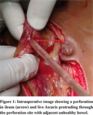
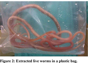
A young adult male (25 years) presented in our emergency department with acute onset severe pain abdomen for 48 hours without any radiation or relief with symptomatic medications. He denied any past history suggestive of gastric or duodenal ulcer. There was also no history of analgesic abuse or any trauma. He gave a history of passages of worms in faeces a few days back. The patient was febrile on admission with tachycardia, hypotension, and low urine output. The abdomen was tender with guarding and rebound tenderness. On admission, the white blood cell count was 16600/ml. His blood urea nitrogen and creatinine were 58.1 mg/dl and 1.1 mg/dl, respectively. Abdominal X-ray revealed gas under the dome of the diaphragm. After fluid resuscitation, a laparotomy was performed. A perforation was found in the ileum (30 cm proximal to the ileocaecal junction) and a live motile worm was seen coming out through the perforation site (Figure 1) and wandering free intothe peritoneum. Few live worms could be palpated within the adjacent unhealthy ileum. He underwent ileal resection with end ileostomy with extraction of worms (Figure 2). He recovered uneventfully except for surgical site infection which was managed conservatively. The histopathology report showed non-specific inflammation without any evidence of a granuloma. He was started on anti-helminthic drugs. Ileostomy was closed after 3 months and he is symptom-free at 3 years follow-up.
Discussion
Ascariasis is still prevalent in some countries and it causes about 10,00,000 new cases and 60,000 deaths in a year worldwide.2,3 Adult worms stay in the lumen of the small bowel and usually cause chronic symptoms like vague abdominal pain, nausea, diarrhoea, and malnutrition/anaemia. However, surgical complications are not uncommon, especially in developing countries. Surgical complications like intestinal obstruction are more common in paediatric and young adults. The cause may be a narrower bowel lumen in children that gets occluded by a worm bolus more easily than an adult with a wider circumference of the bowel. In endemic areas, it has been shown that between 5-35% of cases of bowel obstruction are due to ascariasis.1 There are a few steps involved in the development of ascariasis-induced bowel obstruction. Firstly, worms form a large bolus that can cause mechanical intestinal obstruction. Secondly, intussusception may be caused by a worm bolus. Thirdly, worm-loaded bowel may get twisted and cause volvulus, sometimes leading to bowel ischemia and perforation. Lastly, the inflammatory response against worm-related toxins may cause bowel obstruction.
Though obstruction is common, perforation of theintestine due to helminthic infestation is rare. Different sites of bowel perforation have been described in literature like the stomach7, duodenum8, jejunum9, ileum1 and colon10. Pathologically, perforation by Ascaris worms can be of two types: primary and secondary. In primary perforation, the bowel is healthy, while secondary perforation is related to pre-existing conditions like peptic ulcer, typhoid fever, and tubercular or amoebic ulcer.5 In primary perforation, it has been thought that worm secretes some lytic material and this is associated with screwing movement of the worms, leading to perforation of the naturally impassable intestinal wall.5,6
Despite improvement in sanitation measures and consciousness regarding hygiene, helminthic infestations are still common in some part of India. Habits of consuming properly cleaned food, use of sanitary toilets, avoiding barefoot walking, enforcing laws to maintain hygiene, and health programs to diagnose and treat chronic carriers should be established. Additionally, routine anti-helminthic chemoprophylaxis programsfor all preschool and school-going kids may decrease the possibility of worm infestation and associated surgical complications like obstruction or perforation.
Conclusion
In conclusion, ascariasis-related obstruction and perforation should be taken into consideration as an alternate diagnosis of surgical acute abdomen especially in paediatric population and young adults in endemic areas. Early diagnosis and prompt treatment can reduce surgical complications and morbidity.
References
- Mishra P K, Agrawal A, Joshi M, et al. Intestinal obstruction in children due to Ascariasis: A tertiary health centre experience. Afr J PaediatrSurg2008;5:65-70
- Drake L, Bundy D. Multiple helmith infections in children: Impact and control. Parasitological 2001;122:73-81.
- Cooper PJ, Chico ME, Sandoval C, et al. Human infection with Ascaris lumbricoides is associated with a polarized cytokine response. J Infect Dis 2000;182:1207-13.
- Vilamizar E, Mendez M, Bonilla E, Varon H, et al. Ascaris lumbricoides infestation as a cause of intestinal obstruction in children: Experience with 87 cases. J PediatrSurg1996;31:201-5.
- Singh S, TayalA, Kaur V.Ascaris lumbricoides and duodenal perforation: a case report. JIMSA. 2011;24(4):181-2
- Rao PL, Shenoy MG, Venkatesh A, et al. Intraperitoneal round worm abscess. Indian Pediatr. 1980;17:633-6
- Gupta S, Kumar S, Satapathy A, et al. Ascaris lumbricoides: an unusual aetiology of gastric perforation. J Surg Case Rep. 2012 Dec 4(11):rjs008.
- Refeidi A. Live Ascaris lumbricoides in the peritoneal cavity. Ann Saudi Med. 2007;27(2):118-121.
- Tejareddy KV, Venkannababu A, Deepakvarma K, et al. Ascaris lumbricoides causing Jejunal Perforation after trivial trauma. Int Surg J 2017;4:3169-3171.
- Molina GA, Torres AR, Llerena PS, et al. Ascaris lumbricoides and its almost deadly complication. J Surg Case Rep. 2018;2018(10):rjy262.