48uep6bbphidcol2|ID
48uep6bbphidvals|1924
48uep6bbph|2000F98CTab_Articles|Fulltext
Case Report
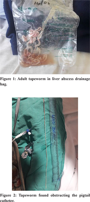
A 55 yr old, a chronic alcoholic, non diabetic, immunocompetent male came with history of high grade fever and right upper quadrant abdominal pain, non colicky and constant in nature for the last 2 weeks. He was non vegetarian by diet. On evaluation found to have two liver abscess, first one of size 9.5x14x10.8 cm in segment VIII and the other one of size 8.1x 7.4x 7.7 cm in segment V of right lobe of liver. His haemoglobin was 8.5 gm/dl, WBC count of 16,600/cumm (N87%, L08%, E 0%), Total Bilirubin was 1.8mg/dl, SGOT/SGPT-146/108 U/L and Alkaline phosphatase was 231 U/L(56-119 U/L).Serum IgE 612.8(0-150). He was started on broad spectrum antibiotics and USG guided pigtail catheter was kept inside both liver abscess cavity. Blood and liver abscess culture did not yield any growth. He also had right pleural effusion which was drained and culture showed growth of Pseudomonas for which antibiotics were started according to culture and sensitivity. Inspite of above measures fever persisted. After few days, a large adult tapeworm- Taenia solium ( approx 250 cm in length) was noted in one of the abscess drain (Figure.1). It was confirmed by microbiologist. Albendazole therapy was started. After few days, there was no more fluid collection in the drain. CECT abdomen did not show any evidence of biliary enteric fistula. After 3 days, when the drain was removed, one more tapeworm ( length approx 35 cm ) (Figure 2) was recovered obstructing the pigtail catheter. Further, no more worms recovered. He improved significantly and was discharged.
Discussion
Taenia solium is the pork tapeworm belonging to cestodes. The adult worm is found in humans. It is white in color, flat and ribbon like body and 2 to 3 m in length. The main body, the strobila, consists of a chain of segments known as proglottids. Each proglottid is a reproductive unit. It completes its life cycle in humans as the definitive host and pigs as intermediate host. It is transmitted to pigs through human feces and to humans through uncooked or under cooked pork. Pigs ingest embryonated eggs called morula which develop into larvae, the oncospheres, and ultimately into infective larvae, cysticerci. A cysticercus grows into an adult worm in human small intestine. Humans are colonized by the larval stage, the cysticercus. A cysticercus is oval in shape, containing an protoscolex, which everts once the organism is inside the small intestine. Using the scolex, it anchors to the intestinal wall. Its strobila lengthens as new proglottids are formed at the neck. About 10 weeks after initial colonization, it becomes an adult worm. As a hermaphrodite, it reproduces by self fertilization, as hermaphrodite or cross fertilization.1,2
Taenia saginata and T. solium are difficult to differentiate by parasitological examination because their eggs are indistinguishable. Differentiation of the two human Taenia species is based on the number of uterine branches present in well-preserved gravid proglottids or on the absence or presence of hooks in the scolex of the tapeworm.3 Taenia solium usually involves the central nervous system but other organs like the heart, skeletal muscle and the orbit can also be involved. Rarely, the liver can also be the site of involvement. In its complete life cycle, neither adult T. solium nor saeginata never dwells the biliary system or inhabit the liver. A rare case of hepatic cysticercosis was reported by Satyanarayan et al.4 They confirmed hepatic cysticercosis by ultrasound imaging of abdomen which showed multiple cysticerci with scolices and IgG of cysticercosis detected by ELISA was also strongly positive. To the best of my knowledge, this is the first case report which showed the presence of adult Taenia solium in liver abscess fluid. We have also done CECT abdomen to rule out the presence of any biliary enteric fistula. It was not clear weather tapeworm was the cause of liver abscess or a bystander. Both the worms were present in only one of the abscess cavity. The other larger abscess did not show the presence of adult tapeworm and both the abscess were resolved after prolong course of antibiotics. So probably the abscesswere pyogenic and secondarily infected by tapeworm.
References
- Carter, Burton J. Bogitsh, Clint E. (2013). Human Parasitology (4th ed.). Amsterdam: Academic Press. pp. 241–244.
- Gutierrez, Yezid (2000). Diagnostic Pathology of Parasitic Infections with Clinical Correlations (2nd ed.). New York [u.a.]: Oxford University Press. pp. 635–652.
- Rausch R L. Parasitology: retrospect and prospect. J Parasitol. 1985;53: 484–491.
- Vishwanath Sathyanarayanan, CharuduttSambhaji, KavithaSaravu, AbdulRazak, AshwinPolnaya, SNRao. A rare case of hepatic cysticercosis. Asian Pacific Journal of Tropical BiomedicineVolume 1, Issue 1, Supplement, September 2011, Pages S139-S140.