48uep6bbphidvals|351
48uep6bbph|2000F98CTab_Articles|Fulltext
Laparoscopic cholecystectomy is now accepted as the “gold standard” for treatment of biliary pancreatitis to prevent further attacks.[1] When dissection of the Calot’s triangle is difficult, partial cholecystectomy has been proposed as a safer operation.[2] The remnant stump, which includes the Hartmann’s pouch, should be cleared of residual gall stones at the time of surgery.[3] However, some of these remnants may retain stones,or form new ones in the post operative period.[3,4] These patients may present with “post cholecystectomy pain” at varying intervals from the primary surgery due to the residual or recurrent stones.[5] The remnant gall bladder and cystic duct stump must be kept in mind while evaluating these patients, especially if a history of partial cholecystectomy is available.[5]We present four such patients who presented with post cholecystectomy biliary pancreatitis, with a view to highlight this problem.
Case Report
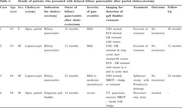
The patients details are provided in Table 1. There was one female and three males with a mean age of 56.6 years (range 45- 69 years). Two patients had undergone open partial cholecystectomy and two, laparoscopic cholecystectomy as the index operation. The details of laparoscopic surgery done (complete/ partial) and the indication for partial cholecystectomy as done in the two open cases were not available. The time of presentation after primary surgery ranged from twelve to twenty-four months. All these patients had a residual gall bladder stump detected on imaging. The gall bladder was detected on Ultrasonography (USG) in one patient, Magnetic Resonance Cholangio Pancreatography (MRCP) showed sludge in the remnant in two patients. Computerized Tomography (CT) was used only in one patient with necrotizing pancreatitis. Endoscopic Ultrasonography (EUS) showed the remnant gall bladder with stones in two patients. In all the patients, the common bile duct did not reveal stones or sludge.
Two of these patients had severe pancreatitis. One patient presented with a pseudocyst of the pancreas and was treated with sphincterotomy and pseudocyst drainage. Two patients who presented with mild pancreatitis were treated with excision of the stump. None of the patients who underwent stump excision or sphincterotomy has had a further attack (mean follow up 28 months).

Discussion
In laparoscopic cholecystectomy, the cystic duct is divided close to the gall bladder to avoid bile duct injury, leading to a longer cystic duct remnant compared to open cholecystectomy.[6] In patients who undergo a partial cholecystectomy, in addition to the cystic duct remnant, a portion of the gall bladder is left behind, to avoid Calot’s triangle dissection.[3,7] Cystic duct remnant, defined as a residual duct or gall bladder remnant greater than 1cm in length, in the presence of stones, can cause post-cholecystectomy syndrome and complications including biliary pancreatitis.[8] The incidence of PCS is reported to be between 10-40%.[5,9]
Routine imaging such as USG may not be able to detect the remnant stump, as was the case in two of our patients, due to the small size, unless gross dilatation of the stump has taken place or a large filling defect is clearly visualized.[5] Hence, when patients who have undergone laparoscopic or open partial cholecystectomy present with biliary pancreatitis, it is advisable to use modalities like MRCP and EUS, along with ERCP and sphincter of Oddi (SOD) manometry.[1,4,5] Both MRCP and EUS have been found to be useful in imaging the remnant stump. [6,8,10,11,12] The differential diagnosis of a cystic lesion in the extra hepatic biliary tree, after cholecystectomy, includes a gall bladder remnant, secondary dilatation of the cystic duct stump, gall bladder duplication and type II choledochal cyst.[4]
In patients who have biliary pancreatitis after cholecystectomy, the differential diagnosis includes residual or recurrent bile duct stones, cystic duct stones, remnant gall bladder stones and SOD dysfunction. EUS has been shown to be accurate for the identification of gallbladder sludge, common bile duct stones, and pancreatic disease. ERCP and sphincter of Oddi manometry should generally be reserved for patients with multiple unexplained attacks and negative EUS results, who have previously undergone cholecystectomy.[13,14,15] The treatment option in case of recurrent pancreatitis following a previous cholecystectomy could be an endoscopic sphincterotomy or resection of the remnant. There are increasing numbers of reports favouring resection of the residual cystic duct/ gall bladder remnant in patients suffering from post cholecystectomy syndromes.[6,8] Completion cholecystectomy or “recholecystectomy” is now gaining importance as the definitive treatment for residual gall bladder remnant and can be performed laparoscopically when feasible.[1,10,16,17]
However, resurgery can be technically challenging. In our patients, the two who underwent completion cholecystectomy had demonstrable stones in the remnant stump. Neither of them has had a recurrence of symptoms after removal of the residual gall bladder. A sphicterotomy was performed in the patient who had sludge seen in the remnant on MRCP as this patient had already undergone pseudocyst drainage after the laparoscopic cholecystectomy and we thought it would be difficult to approach the stump surgically. There are some conceptual advantages of remnant excision over sphincterotomy. Besides eliminating other problems associated with remnant stones (e.g. post cholecystectomy pain), it avoids the side effects as well as the long term recurrent stenosis associated with a sphincterotomy.[18]
In conclusion, in patients who have undergone partial cholecystectomy for biliary pancreatitis, recurrence of the pancreatitis may be due to stones in the remnant gall bladder.
References
1. Chowbey PK, Bandyopadhyay SK, Sharma A, Khullar R, Soni V, Baijal M. Laparoscopic reintervention for residual gallstone disease. Surg Laparosc Endosc Percutan Tech. 2003;13:31–5.
2. Chowbey PK, Sharma A, Khullar R, Mann V, Baijal M, Vashistha A. Laparoscopic subtotal cholecystectomy: a review of 56 procedures. J Laparoendosc Adv Surg Tech A. 2000;10:31–4.
3. Philips JA, Lawes DA, Cook AJ, Arulampalam TH, Zaborsky A, Menzies D, et al. The use of laparoscopic subtotal cholecystectomy for complicated cholelithiasis. Surg Endosc. 2008;22:1697–700.
4. Mergener K, Clavien PA, Branch MS, Baillie J. A stone in a grossly dilated cystic duct stump: a rare cause of post cholecystectomy pain. Am J Gastroenterol. 1999;94:229–31.
5. Hassan H, Vilmann P. Insufficient cholecystectomy diagnosed by endoscopic ultrasonography. Endoscopy. 2004;36:236–38.
6. Lum YW, House MG, Hayanga AJ, Schweitzer M. Postcholecystectyomy syndrome in the laparoscopic era. J Laparoendosc Adv Surg Tech A. 2006;16:482–85.
7. Beldi G, Glattli A. Laparoscopic subtotal cholecystectomy for severe cholecystitis. Surg Endosc. 2003;17:1437–9.
8. Shaw C, O’Hanlon DM, Fenlon HM, McEntee GP. Cystic duct remnant and the ‘post cholecystectomy syndrome’. Hepatogastroenterology. 2004;51;36–8.
9. Venu RP, Geenen JE. Post cholecystectomy syndrome. In: Yamada T, editors. Textbook of gastroenterology. 2nd ed. Philadelphia: Lippincott; 1995.p.2265–77.
10. Demetriades H, Pramateftakis MG, Kanellos I, Angelopoulos S, Mantzoros I, Betsis D. Retained gall bladder remnant after laparoscopic cholecystectomy. J Laparoendosc Adv Surg Tech A. 2008;18:276–9.
11. Petrone MC, Arcidiacono P, Testoni PA. Endoscopic ultrasonography for evaluating patients with recurrent pancreatitis. World J Gastroenterol. 2008;14:1016–22.
12. Topazian M, Salem RR, Robert ME. Painful cystic duct remnant diagnosed by Endoscopic Ultrasound. Am J Gastroenterol. 2005;100:491–5.
13. Wilcox CM, Varadarajulu S, Eloubeidi M. Role of endoscopic evaluation in idiopathic pancreatitis: a systematic review. Gastrointest Endosc. 2006;63:1037–45.
14. Coyle WJ, Pineau BC, Tarnasky PR, Knapple WL, Aabakken L, Hoffman BJ, et al. Evaluation of unexplained acute and acute recurrent pancreatitis using endoscopic retrograde cholangiopancreatography, sphincter of Oddi manometry and endoscopic ultrasound. Endoscopy. 2002;34:617–23.
15. Scheiman JM, Carlos RC, Barnett JL, Elta GH, Nostrant TT, Chey WD, et al. Can endoscopic ultrasound or magnetic resonance cholangiopancreatography replace ERCP in patients with suspected biliary disease? A prospective trial and cost analysis. Am J Gastroenterol. 2001;96:2900–4.
16. Walsh RM, Ponsky JL, Dumot J. Retained gallbladder/ cystic duct remnant calculi as a cause of post cholecystectomy pain. Surg Endosc. 2002;16:981–4.
17. Palanivelu C, Rangarajan M, Jategaonkar PA, Madankumar MV, Anand NV. Laparoscopic management of remnant cystic duct calculi: a retrospective study. Ann R Coll Surg Engl. 2009;91:25–9.
18. Rolny P, Andrén-Sandberg A, Falk A. Recurrent pancreatitis as a late complication of endoscopic sphincterotomy for common bile duct stones: diagnosis and therapy. Endoscopy. 2003;35:356–9.