|
|
|
|
 |
|
| |
 |
|
|
Paediatric Gastroenterology |
|
|
|
|
|
Keywords :
antral gastritis, Helicobacter pylori, gastric atrophy, childhood |
|
|
S Boukthir, 1S Mazigh Mrad, 1 N Kalach2 , A Sammoud1
Service de Médecine Infantile C Hôpital d’Enfants,
Bab Saadoun 1007 Tunis Jebbari,
Tunisia.1
Clinique de Pédiatrie Saint Antoine Hôpital Saint Vincent de Paul Université Catholique,
Bd de Belfort 59020 Lille,
France2
Corresponding Author:
Dr. Samir Boukthir
Email: aboukthir@yahoo.fr
DOI:
http://dx.doi.org/
Abstract
Aim: To assess the prevalence of gastric atrophy (GA) in Tunisia (a high prevalence region for Helicobacter pylori), and describe its histological, clinical and endoscopic features in children.
Methods: 345 children, 151 male and 194 female, mean age 8.6 ± 3.7 years, underwent upper gastrointestinal (UGI) endoscopy with gastric biopsies for recurrent abdominal pain (n=232, 67.2%), vomiting (n=72, 20%) associated with or without upper gastrointestinal bleeding (n=59, 17.1%) and miscellaneous causes (n=53, 15.4 %). Biopsies performed both in the gastric antrum (n=2) and corpus (n=2) were analysed for histological assessment according to the updated Sydney classification system and bacterial culture. A positive result was recorded where histology and/or culture were positive, confirming the presence of H.pylori infection (H. pylori +ve). A negative result was recorded when both tests were concomitantly negative (H. pylori -ve).
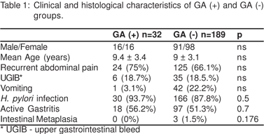
Results: 9.3% (32/345) of the total population, and 14.5% (32/221) of chronic gastritis patients exhibited GA, M/F: 16/16, mean age (SD) 9.4 (3.4) years. Amongst the 32 children with GA, 30 (93.7%) were H. pylori +ve and 2 (6.3%) were H. pylori -ve. GA was localised in the antrum (n=26, 81.2%), the fundus (n=2, 6.3%) and was also seen in both (n=4, 12.5%). GA was categorised as mild, grade 1 (n=18, 56.3%); moderate, grade 2 (n=13, 46.6%); and severe, grade 3 (n=1, 3.1%). GA was associated with mild active gastritis in 18 cases (56.3%). The prevalence of moderate or severe antral GA was detected in 9/26 (34.6%) of H. pylori +ve vs. any of H. pylori -ve ( p=0.4), whereas GA in the corpus was detected in 1/2 (50%) vs. none, respectively. None exhibited intestinal metaplasia. There were no clinical features specific to this pathology. UGI endoscopy in GA patients showed nodular gastritis (n=17, 53.1%), congestive gastritis (n=9, 28.1%), and normal tissue (n=6, 18.8%). GA was significantly associated with H. pylori infection (p<0.0001) and nodular gastritis (p<0.005).
Conclusion: GA was found in 9.3% of Tunisian children undergoing UGI endoscopy andwas significantly associated with H. pylori infection and nodular gastritis.
|
48uep6bbphidvals|241 48uep6bbphidcol2|ID 48uep6bbph|2000F98CTab_Articles|Fulltext Helicobacter pylori (H. pylori) infection is one of the main causes of gastric atrophy (GA) in adults. H. pylori is typically acquired in childhood, yet little is known about the prevalence of GA in childhood because it is not systematically sought during upper gastrointestinal (UGI) endoscopy.Tunisia is a high prevalence zone for H. pylori infection. This study aims at assessing the prevalence of GA and describes the histological characteristics, and clinical and endoscopic features in Tunisian children.
Methods
A retrospective analysis of the endoscopic files during the last 5 years revealed that 345 children, 151 male and 194 female, mean age 8.6 ± 3.7 years (range 1-18 years), underwent UGI endoscopy with gastric biopsy for recurrent abdominal pain (n=232, 67.2%), vomiting (n=72, 20.9%) associated with or without upper gastrointestinal bleeding (UGIB) (n=59, 17.1%) and miscellaneous causes (n=53, 15.4%). Biopsies obtained from the gastric antrum (n=2) and corpus (n=2) were analysed for histology, according to the updated Sydney classification[1] and the tissue obtained was cultured for bacteria according to a previously prescribed method.[2] The updated Sydney System three-tiered visual analogue scales were used to grade the severity of gastric atrophy, which is categorised as mild, moderate and severe.[1] The results of the histological analysis and bacterial culture of the gastric biopsy specimens were carried out blindly, without knowing the results of any other examination. A positive result was retained when histology and/or culture were positive, confirming the presence of H. pylori infection (H. pylori +ve). A negative result was retained when both tests were concomitantly negative (H. pylori -ve). Statistical quantitative parameters were analysed by the Chi square (Chi2) and the exact Fischer tests. A p value of < 0.05 was considered significant.
Results
H. pylori +ve and chronic gastritis, respectively, were detected in 215/345 (62.3%) and 221/345 (64.04%) children, sex ratio M/F: 0.89 and 0.93; mean age (SD) 91 (45) and 109 (43) months.
9.3% (32/345) of the total population, 14.5% (32/221) of chronic gastritis patients exhibited GA, M/F: 16/16, mean age (SD) 9.4 (3.4) years. Among the 32 children with GA, 30 (93.7%) were H. pylori +ve and 2 (6.3%) H. pylori –ve.
Children between 12 and 18 years showed significantly higher prevalence of GA compared to children between 6 and
12 years (53% vs. 31%) (p < 0.05). GA was localised in the antrum (n=26, 81.2%), the fundus (n=2, 6.25%) and was seen in both (n=4, 12.5%). GA was grouped as mild, grade 1 (n=18, 56.25%); moderate, grade 2 (n=13, 46.62%); and severe, grade 3 (n=1, 3.12%). GA was associated with mild active gastritis in 18 cases (56.25%). The prevalence of moderate or severe antral GA was detected in 9/26 (34.6%) of H. pylori +ve versus any of H. pylori –ve, p=0.4, whereas GA in the corpus was detected in 1/2 (50%) vs. none, respectively. None exhibited intestinal metaplasia. There was no clinical feature specific to the trait under study (Table 1). Investigation of those patients ruled out pernicious anaemia, celiac disease and Crohn’s disease. UGI endoscopy in GA patients showed nodular gastritis (n=17, 53.1%), congestive gastritis (n=9, 28.1%), and normal tissue (n=6, 18.8%). GA was significantly associated with H. pylori infection (p<0.0001) and nodular gastritis (p<0.005). H. pylori +ve patients showed significantly higher prevalence of chronic GA and activity of gastritis, compared with H. pylori –ve patients (p<0.00001).
Discussion
Chronic H. pylori infection is one of the main causes of chronic gastritis. Its prevalence in asymptomatic Tunisian children is 30.4%3 and reaches 54% in children with recurrent episodes of unexplained abdominal pain.[4] GA is a multifocal pangastritis, involving independent foci in the corpus and antrum of the stomach.[5] Several clinical reports confirm that GA is a pathology not limited to adult patients.6,7 Its frequency in children is however unknown because it is not systematically sought during UGI endoscopy.[7] In a study conducted among 173 children from countries with high gastric cancer incidence; 58 from Korea and 115 from Colombia, Ricuarte8 et al reported H. pylori in 85% of Colombian children vs. 17% of Korean children (p<0.01). Atrophic mucosa near the antrum-corpus border was present in 16% of children.
In a retrospective study including 196 patients, ages 1 through 16 years [H. pylori +ve (n=131), H. pylori –ve (n=65)] Kato[9] et al reported that H. pylori-induced gastric inflammation can cause atrophy in Japanese children, predominantly in the antrum. The prevalence of grade 2 or 3 atrophy in the antrum was 10.7% in H. pylori-infected patients and 0% in the noninfected patients (p < 0.01) and in corpus 4.3% and 0%, respectively (p = 0.20). In the antrum, atrophy was significantly correlated with activity, whereas in the corpus, atrophy correlated with H. pylori density, inflammation, and activity.[9]
Intestinal metaplasia associated with H. pylori infection has already been reported in children. [6] In contrast with chronic gastritis, none of the patients with GA exhibited intestinal metaplasia. Kato et al[9] showed that the frequency of intestinal metaplasia in the H. pylori-infected and in the non-infected groups was 4.6% and 4.6% in the antrum and 0% and 4.2% in the corpus, respectively. It is suggested that atrophy exists in children, since atrophy usually precedes intestinal metaplasia in adults. A large number of B-lymphocytes and apoptosis in the surface epithelium are seen in patients with H. pylori infection and may be related to the development of atrophy and intestinal metaplasia.[6]
The prevalence of GA seems to increase with advancing age in H. pylori-infected patients, compared to H. pyloriuninfected patients.[10] In a 10-year prospective follow up study, the cumulative progression rate of GA due to H. pylori infection was 6% after 2 years, 22% after 4 years, 34% after 6 years and 43% after 10 years. These atrophic changes were related to neutrophil infiltration caused by H. pylori infection.[11]
The prevalence of GA seems to increase with advancing age in H. pylori-infected patients, compared to H. pyloriuninfected patients.10 In a 10-year prospective follow up study, the cumulative progression rate of GA due to H. pylori infection was 6% after 2 years, 22% after 4 years, 34% after 6 years and 43% after 10 years. These atrophic changes were related to neutrophil infiltration caused by H. pylori infection.[11]
Since GA and intestinal metaplasia have traditionally been considered markers for premalignancy,[7] it remains to be determined whether H. pylori-infected children with gastric
atrophy are at increased risk for gastric cancer.
Conclusion
Gastric atrophy was found in 9.3% of children undergoing upper gastrointestinal endoscopy and was significantly associated with Helicobacter pylori infection and nodular gastritis. Gastric atrophy has to be systematically sought in children, specifically in zones of high Helicobacter pylori prevalence.
References
1. Dixon MF, Genta RM, Yardley JH, Correa P. Classification and grading of gastritis. The update Syndey System. International workshop on the histopathology of gastritis. Houston 1994. Am J Surg Pathol. 1996;20:1161–81.
2. Kalach N, Bergeret M, Benhamou PH, Dupont C, Raymond J. High levels of resistance to metronidazole and clarithromycin in childhood Helicobacter pylori strains. J Clin Microbiol. 2001;39:394–7.
3. Maherzi A, Bouaziz Abed A, Fendri C, Oubich F, Koubaa C, Fauchere JL, et al. [Helicobacter pylori infection: prospective study for asymptomatic Tunisian children] [Article in French]. Arch Pediatr. 2003;10:204–7.
4. Maherzi A, Fendri C, Ben Jilani S, Bousnina S. [Symptomatic Helicobacter pylori infection: prospective study of epidemiological, diagnostic and therapeutic aspects in children in Tunisia] [Article in French]. Arch Pediatr. 1996;3:329–34.
5. Kapadia CR. Gastric atrophy, metaplasia, and dysplasia: a clinical perspective. J Clin Gastroenterol. 2003;36(5 Suppl):S29–36.
6. Guarner J, Bartlett J, Whistler T, Pierce-Smith D, Owens M, Kreh R, et al. Can pre-neoplastic lesions be detected in gastric biopsies of children with Helicobacter pylori infection? J Pediatr Gastroenterol Nutr. 2003;37:309–14.
7. Dimitrov G, Gottrand F. Does gastric atrophy exist in children? World J Gastroenterol. 2006;12:6274–9.
8. Ricuarte O, Gutierrez O, Cardona H, Kim JG, Graham DY, El-Zimaity HM. Atrophic gastritis in young children and adolescents. J Clin Pathol. 2005;58:1189–93.
9. Kato S, Nakajima S, Nishino Y, Ozawa K, Minoura T, Konno M, et al. Association between gastric atrophy and Helicobacter pylori infection in Japanese children: a retrospective multicenter study. Dig Dis Sci. 2006;5199–104.
10. Ohkuma K, Okada M, Murayama H, Seo M, Maeda K, Kanda M, et al. Association of Helicobacter pylori infection with atrophic gastritis and intestinal metaplasia. J Gastroenterol Hepatol. 2000;15:1105–12.
11. Sakaki N, Kozawa H, Egawa N, Tu Y, Sanaka M. Ten-year prospective follow-up study on the relationship between Helicobacter pylori infection and progression of atrophic gastritis, particularly assessed by endoscopic findings. Aliment Pharmacol Ther. 2002;16 Suppl 2:198–203.
|
|
|
|
|
|