48uep6bbphidvals|347
48uep6bbph|2000F98CTab_Articles|Fulltext
Eosinophilic enteritis (EE) is a rare disease that is characterized by tissue eosinophilia. The clinical signs and symptoms depend on the layer of the gut predominantly involved. Klein and Tally et al. have classified this disorder into mucosal, submucosal and subserosal disease, based on the layer of the gut predominantly involved.[1,2,3] The most prevalent form is the one with predominant involvement of the mucosal layer with symptoms of colicky abdominal pain, nausea, vomiting, diarrhoea and weight loss. The exact pathogenesis of the disease remains unknown but, at least in some of these patients with predominant involvement of the mucous layer, high serum IgE levels has been documented. Most patients will have history of food allergy or family history of allergy.[4]
Case Report
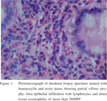
A six year old female child born of non consanguineous parents was admitted with a history of abdominal distension with vague pain and loose stools of two months duration. She had 6-8 loose motions per day associated with borborygmi with occasional vomiting. She had loss of appetite with low grade intermittent fever. There was no history of contact with open case of tuberculosis, drug intake, or worm infestation. Physical examination revealed grade I protein malnutrition, mild anemia and pedal edema. Abdomen was distended with a doughy feel. Investigations revealed a total count of 14,800 cells/cu mm, differential count of 20% polymorph, 28% lymphocytes, 52% eosinophils. Hemoglobin was 9 gm% and peripheral smear showed hypochromic microcytic anemia. Stool examination showed no ova or cysts. Occult blood was negative. Serum albumin was 3 gm/dl. Chest X ray was normal. HIV test and mantoux tests were negative. Ultrasound (USG) abdomen revealed minimal free fluid with thickened bowel wall. Barium meal follow through showed thickened bowel wall with features suggestive of malabsorption. Upper gastrointestinal endoscopy showed no visible abnormality and duodenal biopsy of the mucosa from the 3rd part of duodenum was done. Histopathology showed partial villous atrophy, intra epithelial infiltration with lymphocytes and dense tissue eosinophilia of more than 50/HPF (Figure 1). She had elevated serum IgE levels (196 IU/l). A final diagnosis of eosinophilic enteritis was made. Our patient had involvement of mucosal and subserosal involvement as she had diarrhoea and ascites. . The child was started on oral prednisolone 2mg/kg and tapered over a period of three months. The child showed dramatic response to oral steroid therapy. The child improved clinically with reduction in diarrhoeal frequency and gain in weight. Currently the child is doing well.

Discussion
Chronic diarrhoea in children encompasses a wide variety of causes. When our patient presented with diarrhoea for two months duration, the initial evaluation was carried to rule out some of the conditions peculiar to tropical countries like giardiasis, tuberculosis and persistent infectious diarrhoea. She had peripheral eosinophilia. Upper gastroscopy with duodenal biopsy confirmed the diagnosis of EE. Elevated serum IgE is seen more often in paediatric population than adults with EE.[5] Eosinophilic enteritis is an uncommon disorder that has been documented mainly in adulthood3 and characterized by infiltration by eosinophils of gastrointestinal tract. Patients with mucosal involvement present with nausea, vomiting, diarrhoea and weight loss. Muscular involvement usually presents with intermittent obstructive symptoms or with complications like perforation. Serosal involvement presents with ascites. Rarely, other organs like pleura, pericardium, gall bladder, biliary tree and liver can be affected. The exact etiology is not known although few of these patients may have food allergy or family history of allergy. Canine hookworm, Ankylostoma infestation, drugs like carbamazepine, cotrimoxazole and heavy metals like gold have been incriminated as causes in enterocolitis with tissue eosinophilia. The defects in mucosal integrity may be responsible for localization of various antigens in gut wall resulting in infiltration of eosinophilia in blood and tissues. Favourable response to steroids suggest the possibility of type I hypersensitivity reaction. Eosinophil through toxic cationic protein (MBP) plays a role in pathogenesis of this disease.[3] Food allergy has been noticed in 50% of cases.[6]
Allergic mechanism has been implicated in most of these patients.[7] A viral etiology has also been suggested in some cases.[8] Peripheral eosinophilia is noticed in 80% of patients.[2]
The diagnosis is established by demonstration of tissue eosinophilia on endoscopic, laparoscopic or surgical specimens. However, the endoscopy may be normal or may show mild erythema to nodularity, thickened mucosal folds or even frank ulcerations. Full thickness surgical biopsies may be needed for diagnosis if the disease process is confined to the muscular layer. Ultrasonography, barium contrast studies, computerised tomography of the abdomen may show bowel thickening, nodularity or evidence of gastric outlet obstruction. Stomach is the most common site.[2] Duodenum and rest of the small bowel may be involved.[9] Steroids form the mainstay of therapy and nearly 90% will show favourable response. Mast cell stabilizers like sodium chromoglycate can be used to prevent mast cell degradation and release of histamines and leukotrines. Eosinophilic gastroenteritis in general has good prognosis.
References
1. Klein NC, Hargrove RL, Sleisenger MH, Jeffries GH. Eosinophilic gastroenteritis. Medicine. 1970;49:299–319.
2. Talley NJ, Shorter RJ, Philips SF, Zinsmeister AR. Eosinophilic Gastroenteritis: a clinicopathological study of patients with disease of mucosa, muscular layer and subserosal tissues. Gut. 1990:31:54–8.
3. Talley NJ, Kephart GM, McGovern TW, Carpenter HA, Gleich GJ. Depsosition of eosinophil granule major basic protein in eosinophilic gastroenteritis and celiac disease. Gastroenterology. 1992;103:137–45.
4. Zora JA, O’Connell EJ, Sachs MI, Hoffman AD. Eosinophilic gastroenteritis: a case report and review of the literature. Ann Allergy. 1984;53:45–7.
5. Katz AJ, Goldman H, Grand RJ. Gastric mucosal biopsy in eosinophilic (allegic) gastroenteritis. Gastroenterology. 1977;73:705–9.
6. Scudamore HH, Phillips SF, Swedlund HA, Gleich GJ. Food allergy manifested by eosinophilia, elevated immunoglobulin E level and protein losing enteropathy: the syndrome of allergic gastroenteropathy. J Aller Clin Immunol. 1982;70:129–38.
7. Khan S. Eosinophilic gastroenteritis. Best Pract Res Clin Gastroenterol. 2005;19:177–98.
8. Whitington PF, Whitington GL. Eosinophilic gastroenteropathy in childhood. J Pediatr Gastroenterol Nutr. 1988;7:379–85.
9. Naylor AR. Eosinophilic gastroenteritis. Scott Med J. 1990;35:163–5.