Vidhyachandra Gandhi, Sujith Philip, Amit Maydeo, Nilesh Doctor
Consultant GI & Transplant Surgeon
Department of Surgical Gastroenterology,
20th floor, Jaslok Hospital & Research Centre,
15, Dr. G Deshmukh Marg,
Pedder road, Mumbai – 400026
Corresponding Author:
Dr. Nilesh Doctor
Email: drnileshbela@gmail.com
drgandhivv@gmail.com
48uep6bbphidvals|315 48uep6bbph|2000F98CTab_Articles|Fulltext Subcapsular hematoma of the spleen is a rare hemorrhagic complication of pancreatitis.[1,2,3,4] We report a patient with a large subcapsular splenic hematoma as a complication of recurrent acute pancreatitis. The origin of the hematoma was thought to be rupture of pancreatic pseudocysts in the tail of the pancreas.
Case Report
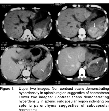
A 35 years male was admitted with abdominal pain, tachycardia, hypotension, pallor and anaemia (Hb – 5.0 gm/ dl). Physical examination revealed fullness and tenderness in the left hypochondrium. Liver function tests were normal, with raised serum amylase (375 U/L) and lipase (633 U/L) levels. He gave history of recurrent episodes of mild acute pancreatitis of two years duration. Three months prior to admission, an ultrasound revealed multiple pancreatic pseudocyts, with no change in size on follow up scans. Celiac angiography showed no evidence of active bleed or formation of pseudoanuerysm. CECT of abdomen revealed pseudocyts within tail of pancreas, splenic hilum and paraesophageal region with large ruptured subcapsular splenic hematoma (Figure 1). He was resuscitated and taken up for emergency laparotomy. On exploration, he had hemoperitoneum, large clots in perihepatic and perisplenic area, a ruptured splenic hematoma and a pseudocyst in the tail of the pancreas. Distal pancreatectomy with splenectomy was done. Postoperative course as well as six month follow up has been uneventful.
Discussion
Proxmitiy of pancreatic tail to the splenic hilum provides a potential for bleeding complications in pancreatitis.[2,3] These could include splenic vein thrombosis, arterial pseudoaneurysms, bleed in a pseudocyst, splenic hematoma and splenic rupture [1,2,3,4]. These complications occur in 1–5% of pancreatitis.[3] The possible mechanisms are: (1) splenic vessel thrombosis and pseudoaneurysm formation; (2) dissection of a pancreatic pseudocyst into the hilum of the spleen, which can cause splenic rupture, splenic infarction, arterial hemorrhage, or venous thrombosis; or (3) extension of the inflammatory process from the tail of the pancreas into the hilum of the spleen, which may induce hematoma formation. [2,3,4]
Splenic hematoma is a rare cause of hemorrhage in pancreatitis and may need urgent surgical intervention. In a large series of 500 patients with chronic pancreatitis, an estimated prevalence of 0.4% was reported.[1] Local factors (thrombosis of the splenic artery or veins, intrasplenic pseudocysts, perisplenic adhesions, enzymatic digestion) and coagulation disorders may play a role in the pathogenesis of splenic hematoma.[4,5] The splenic hematoma in this case was probably caused by extension of the inflammatory process or erosion of pseudocyst from the tail of the pancreas into the splenic hilum. As splenic involvement in patients with acute or chronic pancreatitis is uncommon, the diagnosis of subcapsular hematoma of the spleen needs a high index of suspicion. Patients with pancreatitis presenting with a mass in the left upper quadrant, pain radiating to the left shoulder, and a fall in haemoglobin should be suspected to have a hemorrhagic complication and urgently admitted. Abdominal CT should be performed early. This will help in making a diagnosis of a bleed within a pseudocyst or a pseudo aneurysm or splenic complications. Angiography is indicated if splenic artery pseudoaneurysm, splenic vein thrombosis or active bleeding is suspected.[5,6]
The management of subcapsular splenic hematoma in pancreatitis remains controversial. Surgical treatment with splenectomy, percutaneous drainage, and observation are management options. Kuramitsu et al[6] reported 1 case with chronic pancreatitis and a large subcapsular splenic hematoma (10 × 10 cm). After conservative management for 6 weeks, the size of the hematoma had not change, rupture of the hematoma with peritonitis occurred after a relapse of pancreatitis, which prompted surgical intervention.[6] They suggested that in cases of large splenic hematoma (> 5 cm) as a complication of pancreatitis, pressure reduction by percutaneous drainage or laparotomy should be administered as early as possible. Some reports have advocated aggressive management with early splenectomy to avoid splenic rupture.[1,5] Thompson and Ashley [5] described 3 cases with acute pancreatitis accompanied by large subcapsular hematoma of the spleen. Two patients recovered after splenectomy, but 1 patient died due to continuing blood loss. They suggested that treatment of splenic hematoma should be splenectomy in majority of patients to prevent continuing blood loss and potential rupture.[5] Percutaneous drainage for splenic subcapsular collections may be a feasible treatment, but there are only a few reports.[7,8]
Rypens et al[4] reviewed 16 patients with splenic parenchymal complications due to acute or chronic pancreatitis. Subcapsular splenic collections were detected in 11 patients. Two patients underwent emergency splenectomy, 2 patients received delayed splenectomy, and the others were treated conservatively. Time for healing of splenic lesions varied from 1 week to 4 months. The authors suggested that most splenic complications of pancreatitis regress spontaneously and may be managed conservatively. Surgical indication is based on clinical findings. Patel et al[9] reported a case of large subcapsular splenic hematoma(11.1×9.5 cm) resulting from pancreatitis that was managed conservatively with a good outcome. A CT scan performed 4 months later showed marked resolution of the hematoma. They suggested that it is appropriate to manage splenic subcapsular hematoma conservatively in a hemodynamically stable patient who is showing improving symptoms and signs.
In conclusion, splenic complications of pancreatitis should be recognised early and managed promptly. Surgical intervention may be the therapy of choice for hemodynamically unstable patients.
References
1. Malka D, Hammel P, Lévy P, Sauvanet A, Ruszniewski P, Belghiti J, et al. Splenic complications in chronic pancreatitis: prevalence and risk factors in a medical–surgical series of 500 patients. B J Surg. 1998;85:1645–9.
2. Calvo E, Cigüenza R, Alvarez-Sala JL, Massa B, Espinós D. Spontaneous rupture of the spleen in chronic pancreatitis: an often forgotten complication. Gastroenterol Clin Biol. 1992;16:194.
3. Fishman EK, Soyer P, Bliss DF, Bluemke DA, Devine N. Splenic involvement in pancreatitis: spectrum of CT findings. Am J Roentgenol.1995;164:631–5.
4. Rypens F, Devière J, Zalcman M, Braudé P, Van de Stadt J, Struyven J, et al. Splenic parenchymal complications of pancreatitis: CT findings and natural history. J Comput Assist Tomogr. 1997;21:89–93.
5. Thompson JE Jr, Ashley SW. Subcapsular hematoma of the spleen associated with acute pancreatitis. Surgery. 1997;121:231–3.
6. Kuramitsu T, Komatsu M, Ono T, Nakajima K, Funaoka M, Kato J, et al. Ruptured subcapsular giant hematoma of the spleen as a complication of chronic pancreatitis. Intern Med. 1995;34:564–8.
7. Siu TL. Percutaneous drainage of spontaneous subcapsular haematoma of the spleen complicating chronic pancreatitis. Surgeon. 2004;2:52–5.
8. Vyborny CJ, Merrill TN, Reda J, Geurkink RE, Smith SJ. Subacute subcapsular hematoma of the spleen complicating pancreatitis: successful percutaneous drainage. Radiology. 1988;169:161–2.
9. Patel VG, Eltayeb OM, Zakaria M, Fortson JK, Weaver WL. Spontaneous subcapsular splenic hematoma: a rare complication of pancreatitis. Am Surg. 2005;71:1066–9.
|