Anadi Nath Acharya, Shahana Gupta
Department of Surgery
Institute of Post Graduate Medical
Education and Research & SSKM Hospitals,
Kolkata, India
Corresponding Author:
Dr. Shahana Gupta
E-mail: shahanagupta@yahoo.co.in
Abstract
Seven cases of peritoneal hydatidosis were reviewed. Of these, one had disseminated primary peritoneal echinococcosis, a rare presentation, whereas the rest were secondary to hepatic or splenic lesions. They were treated with a preoperative course of antihelminthics followed by surgery, which consisted of removal of the peritoneal cysts along with de-roofing and omentoplasty for the hepatic lesions and splenectomy for the splenic hydatid. During follow up, all patients were given a three-month course of albendazole and are doing well.
|
48uep6bbphidcol4|ID 48uep6bbphidvals|292 48uep6bbph|2000F98CTab_Articles|Fulltext Peritoneal hydatidosis is an uncommon finding. Secondary peritoneal hydatidosis is more common than primary. Secondary cases are commonly associated with hepatic hydatid cysts. Only a few cases of primary peritoneal hydatidosis have been reported in the literature till date. In this report we review seven cases of peritoneal hydatidosis of which one is a case of primary disseminated peritoneal hydatidosis while the rest are secondary to hepatic and /or splenic lesions.
Methods
The seven cases of peritoneal hydatidosis that we review in this report attended the surgery OPD of SSKM Hospital, Kolkata during the period 2005-2007. All of them underwent thorough clinical examination followed by ultrasonography (USG) and contrast enhanced CT scan (CECT) of the abdomen. An assessment of serum echinococcal antibody was carried out. All patients were given pre-operative albendazole (10 mg/kg body weight) with two week gaps between each course for 3 months. Monthly liver function tests (LFT) were carried out. All patients except one underwent surgical exploration and were followed up with albendazole for 3 months. USG abdomen during follow up did not show any evidence of recurrence.
Review of cases
Case 1: Primary peritoneal echinococcosis
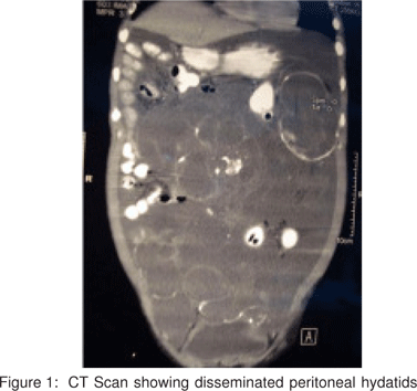
A 48-year-old male patient presented to us with complaints of progressively increasing abdominal distension and vague diffuse abdominal pain for 2 to 3 years’ duration. On examination, there was evidence of free fluid in the abdomen but the rest was normal. He did not have any history of operative intervention. Ultrasonography of the abdomen showed cystic masses throughout the peritoneal cavity resembling hydatid cysts. There were two large cysts approximately 12 cm x 10 cm in size each, one in the pelvis and the other in proximity to the spleen. CECT abdomen confirmed the USG findings (Figure 1). The serum echinococcal antibody titre was raised.
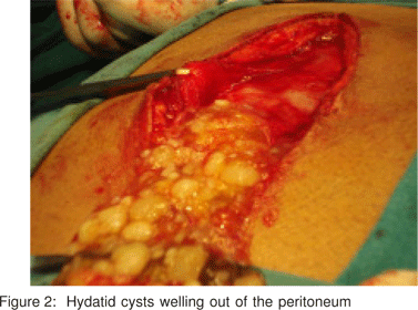
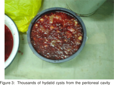
A preoperative diagnosis of primary peritoneal echinococcosis was made. The patient was prescribed albendazole (10 mg/kg body weight) for 3 months with two week gaps between each course. Monthly LFT was performed. Exploration was then carried out. Many thousands of hydatid cysts welled out of the peritoneal cavity as soon as it was opened (Figures 2 and 3). They were disseminated throughout the peritoneal cavity. No hepatic or splenic hydatid was identified. Two large cysts, one in the left hypochondrium, related to the spleen, and the other in the pelvis were found. Many cysts were adherent to the serosa of the small gut and colon, mesentery and mesocolon. Most of the peritoneal cysts were removed and de-roofing and omentoplasty done for the larger cysts. Thorough peritoneal lavage was carried out and the abdomen was closed with drains left in the pelvis on both sides. The postoperative course was uneventful apart from features suggestive of a pelvic collection that settled on conservative therapy. The patient was given albendazole again for three months with two-week gaps between each course. LFT was normal. The patient is doing fine 8 months after surgery. Repeat USG abdomen has not shown any recurrent cysts.


Cases 2 – 7: Secondary peritoneal echinococcosis
The first was a case of hepatic hydatid cyst (in the right lobe) which ruptured following trauma (road traffic accident) leading to secondary peritoneal dissemination. He presented with a history of acute pain abdomen following trauma that settled on analgesic treatment. Echinococcal antibody titre was raised. USG and CECT abdomen confirmed the diagnosis. A course of albendazole was started and planned for surgery. The patient however did not report back for surgery.
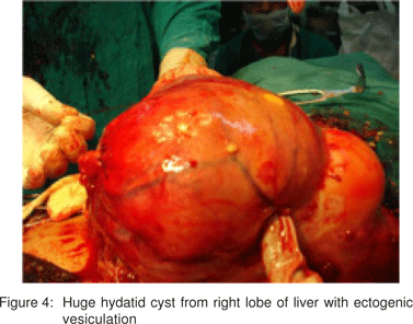
The second was a case of huge hepatic hydatid cyst in a 40-year-old female patient who presented with occasional right upper abdominal pain with an 8 cm x 6 cm lump in the right hypochondrium. CECT abdomen revealed the presence of a huge hepatic hydatid cyst in the right lobe of the liver extending down to the pelvis. The echinococcal antibody titre was raised. After a preoperative course of albendazole, exploration was carried out. A huge hydatid cyst in the right lobe of the liver along with ectogenic vesiculation extending down in to pelvis displacing the pelvic organs was seen (Figure 4). Peritoneal hydatid cysts were also found attached to the greater omentum and serosal surface of the small gut and colon.
The third case, a 38-year-old female patient presented to us with epigastric discomfort. On USG, hydatid cysts were found in the right lobe of the liver as well as in the spleen. On exploration after a course of albendazole, mesenteric and peritoneal hydatids were found, apart from hepatic and splenic hydatids.
Both these patients were treated with de-roofing and omentoplasty for larger cysts, removal of peritoneal hydatids and splenectomy for splenic hydatid cyst. Both were prescribed albendazole for three months postoperatively with two-week gaps between each course. Monthly LFT was performed. The patients are doing well during follow up ten months postoperatively.
The other three cases were of peritoneal echinococcosis following recurrent hydatid cysts of liver diagnosed by USG abdomen and CECT. One of them had undergone percutaneous aspiration, injection and reaspiration (PAIR) procedure and the other two had undergone de-roofing and omentoplasty previously. On re-exploration, small hydatid cysts were seen scattered all over the peritoneal cavity and also adherent to the small bowel serosa, colonic wall, mesentery and mesocolon along with recurrent hepatic lesions in all the cases (Figure 5). De-roofing and omentoplasty for hepatic lesions and removal of the remaining lesions was done. They were prescribed albendazole postoperatively for three months with two-week gaps between each course. Monthly LFT was performed. The patients are all doing fine on follow up three to six months after treatment.

Discussion
Hydatid cyst infection is one of the oldest diseases in humans and animals. It is a zoonotic parasitic disease caused most frequently by Echinococcus granulosus or Echinococcus multilocularis. The life cycle of Echinococcus has a definitive host, commonly dog, and an intermediate host, commonly sheep. Humans are accidental, intermediate hosts infected by coming in to contact with the definitive host or by consuming vegetables or water contaminated by ova.
Apart from the liver, which is the most common organ affected, other organs like the lungs, spleen, kidney, brain, bone or peritoneum may be involved.[1] In contrast to mucosal surfaces, serosal surfaces like pleura or peritoneum constitute a friendly microenvironment for the development of echinococcal cysts.[2,3]
Primary peritoneal echinococcosis is rare.[4,5,6,7,8] Only a few cases have been reported.[9,10,11,12] The mechanism of primary peritoneal infection by the parasite is unknown.
Peritoneal echinococcosis is most commonly secondary to hepatic infection.[13] Microrupture, mostly asymptomatic or leakage of echinococcal fluid during surgery result in peritoneal contamination in 5-10% cases. Post-traumatic microrupture of the hepatic hydatid cyst may also lead to peritoneal seeding. Spontaneous rupture of liver hydatid cysts into the peritoneal cavity is a common complication.
Symptoms due to peritoneal hydatidosis arise commonly from complications due to enlarging abdominal cysts.[9] Rupture into the peritoneum may present as acute abdominal pain. Antigenic fluid released into the peritoneal cavity and absorbed into the circulation may present with acute allergic manifestations.[14] In our series, vague abdominal pain was the most common clinical feature.
Diagnosis is most commonly made through USG or CT scan of the abdomen in which lesions appear as well-defined and circumscribed with or without internal septations. Cysts are staged according to content pattern. Daughter cysts and hydatid sand are also seen, and there maybe associated wall calcification. CT scan is the imaging modality of choice for peritoneal disease.
Several serological tests are used in diagnosis with considerable differences in sensitivity and specificity. Detection of the circulating antigen is less sensitive than antibody detection. ELISA has a sensitivity varying from 64% to 100% depending on the antigen used.[15,16]
Sterilisation of the contents of hydatid cysts by preoperative administration of antihelminthics has been advocated and a concomitant decline in the incidence of recurrence has been reported.[17,18] Preoperative albendazole (10 mg/kg body weight) is administered for 3 to 6 months. There is greater agreement regarding postoperative use of antihelminthics in high-risk patients.[19] The addition of praziquantel to albendazole reportedly produces results superior to those using albendazole alone.[20,21]
Small peritoneal cysts that are asymptomatic may be managed conservatively. Surgical intervention is required for symptomatic and exceptionally large peritoneal cysts. Surgery in such cases may have to be repeated several times to achieve permanent eradication of the disease.[2] Total cystectomy, where possible, is the treatment of choice. When cysts are attached to intraperitoneal viscera, drainage and wide deroofing is safer and is as effective as total cystectomy.[3] With disseminated disease, removal of large symptomatic cysts and of smaller ones as much as possible is advocated. Follow up with antihelminthics is recommended.
References
1. Blumgart LH. Surgery of the Liver, Biliary tract and Pancreas vol.2, 4th ed, W.B. Saunders, Philadelphia, 2007.
2. El Mufti M. In Surgical management of hydatid disease, El Mufti M, editor, London, Butterworth,1989;27-30.
3. El Mufti M. In Surgical management of hydatid disease, El Mufti M, editor, London, Butterworth,1989;31-54.
4. Nadeem N, Khan H, Fatimi S, Ahmad MN. Giant multiple intraabdominal hydatid cysts: case report. J Ayub Med Coll Abbottabad. 2006;18:71-3.
5. Singh RK. A case of disseminated abdominal hydatidosis. J Assoc Physicians India. 2008;56:55.
6. Yadav MK, Mittal P, Rishi JP, Agarwal K. Disseminated abdominalhydatidosis. J Assoc Physicians India. 2007;55:875-6.
7. Vagholkar KR, Nair SA, Rokade N. Bombay Hospital Journal, 2004;46(2); Case Report 13 (http://www.bhj.org/journal/ 2004_4602_april/index.htm).
8. Iqbal SA, Jawaid M, Usmani F. Disseminated Intra-Abdominal Hydatidosis: A Very Rare Presentation. The Internet Journal of Surgery. 2007;11(1).
9. Karavias DD, Vagianos CE, Kakkos SK, Panagopoulos CM, Androulakis JA. Peritoneal Echinococcosis. World J Surg. 1996;20:337-40.
10. Ramji S, Kulshrestha R, Sehgal S, Khandpur SC. Primary peritoneal echinococcosis. Indian Pediatr. 1987;24:258-9.
11. La Torre F, Giacomelli L, Messineti S. Unusual site of hydatidosis: a case with mesenteric location. Minerva Chir. 1988;43:1615-9.
12. Ionescu A, Trufin R, Jakab A, Jutis T. Primary hydatid cyst of the great epiploon with spontaneous rupture: hydatid peritonitis. Rev Chir Oncol Radiol O R L Oftalmol Stomatol Chir. 1985;34:53-6.
13. Wani RA, Malik AA, Chowdri NA, Wani KA, Naqash SH. Primary extrahepatic abdominal hydatidosis. Int J Surg. 2005;3:125-7.
14. Vuitton DA. Echinococcosis and allergy. Clin Rev Allergy Immunol. 2004;26:93-104.
15. Coltorti EA. Standardisation and evaluation of an enzyme immunoassay as a screening test for the seroepidemiology of human hydatidosis. Am J Trop Med Hyg. 1986;35:1000-5.
16. Iacona A, Pini C, Vicari G. Enzyme-linked immunosorbent assay (Elisa) in the serodiagnosis of hydatid disease. Am J Trop Med Hyg. 1980;29:95-102.
17. Morris DL. Preoperative albendazole therapy for hydatid cysts. Br J Surg. 1987;74:805-6.
18. Davidson RN, Bryceson ADM, Cowie AGA, McManus DP, Morris DL. Preoperative albendazole therapy for hydatid cysts. Br J Surg. 1988;75:398.
19. Horton RJ. Chemotherapy of Echinococcus infection in man with albendazole. Trans R Soc Trop Med Hyg. 1989;83:97-102.
20. Taylor DH, Morris DL. Combination chemotherapy is more effective in postspillage prophylaxis for hydatid disease than either albendazole or praziquantel alone. Br J Surg. 1989;76:954.
21. Yasawy MI, Al-Karawi MA, Mohamed AR. Combination of praziquantel and albendazole in the treatment of hydatid disease. Trop Med Parasitol. 1993;44:192-4.
|