48uep6bbphidvals|286
48uep6bbphidcol4|ID
48uep6bbph|2000F98CTab_Articles|Fulltext
Neonatal cholestasic disorders are a group of hepatobiliary diseases occurring within the first 3 months of life, characterized by impairment of bile flow, hyperbilirubinemia, acholic stools and hepatomegaly.[1] Overall, 1:2500 live births is affected with a neonatal cholestatic disorder, with biliary atresia (BA) and idiopathic neonatal hepatitis (NH) accounting for up to 50 to 70% of cases.[2] BA is characterised by complete fibrotic obliteration of the lumen of all or part of the extra hepatic biliary tract. Early surgical treatment offers potential cure in these patients.[3] NH on the other hand is mainly a disease of exclusion and treatment is non-surgical. Surgery in NH can sometimes increase the morbidity. Hence the need to differentiate the two conditions so as to plan early surgery in one and defer surgical intervention in the other. However, there is still no single investigative modality which can convincingly and constantly differentiate one from the other.
Despite clinical improvement after early porto-enterostomy, approximately 70-80% of children with BA eventually require liver transplantation due to biliary cirrhosis leading to endstage liver disease. There is a need to look for ultra-structural liver changes in neonatal cholestatic disorders for any specific diagnostic feature to differentiate BA from NH and also to identify any marker of long term prognosis. This prospective study was initiated to study the electron microscopic features of the liver in patients with BA and NH, to correlate these with light microscopic features and to look for specific differentiating features on electron microscopy.
Material and methods
This study was performed in the Department of Paediatric Surgery. Samples were collected over a period of two years from January 2002 to December 2003 and analysed later. Patients were worked up in the Paediatric Surgical Outpatients’ Department or were referred from Paediatric Medicine for neonatal cholangiopathy. Only patients in whom satisfactory samples were obtained for electron microscopic analysis were included in the study.
Standard liver function tests (LFT) including gamma glutamyl transpeptidase (GGT), TORCH (toxoplasma-rubellacytomegalovirus- herpes) and hepatitis serology, thyroid function tests and prothrombin time were performed in all patients. An ultrasound examination of the abdomen was performed to look for features of BA, to rule out other surgical causes of cholestasis and to look for features of portal hypertension. Mebrofenin hepatobiliary scintigraphy was performed post priming with phenobarbitone to look for hepatocyte function and gut excretion of tracer for 24 hours.[4]
All patients with obstructive jaundice and failure to demonstrate excretion upto 24 hours post mebrofenin hepatobiliary scintigraphy were taken up for minilaparotomy and peroperative cholangiogram (POC) was performed in all patients with a patent gall bladder. Patients with atretic biliary tract on POC and those, in whom POC was not performed due to atretic gall bladder, underwent Kasai’s portoenterostomy. Patients with NH in whom a patent extrahepatic biliary tree was demonstrated on POC, underwent flushing of the biliary tract with saline. Liver biopsy was performed in both BA and NH patients and separate samples were taken for light microscopy and electron microscopic studies. Samples for electron microscopy were collected in 3% glutaraldehyde solution and refrigerated overnight; 1-2 mm slices were made for proper fixation. All samples were processed in the Department of Pathology by the same pathologist.
Results
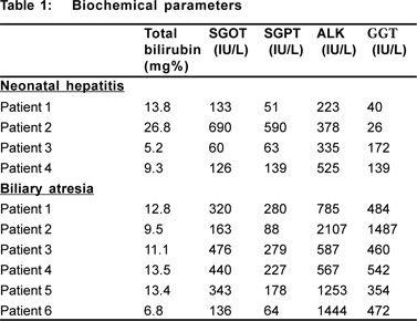
A total of 10 patients with neonatal cholestasis with available tissue specimens for electron microscopic analysis were included in the study. Of the 10 patients, 9 were male and one was female. Six patients had BA while four had NH. Ultrasound examination of the abdomen did not provide for definitive diagnosis in these cases. Mebrofenin hepatobiliary scintigraphy revealed no gut activity in all the patients. Liver function was impaired in all patients with NH and BA although serum alkaline phosphatase and GGT were greatly elevated in BA patients as compared to the NH patients ( Table 1).

The mean age at surgery was 85 days in NH and 110 days in BA. On exploration, the liver appeared to be grossly cirrhotic in three patients with biliary atresia (50%) and these patients had a higher mean age at surgery (153 days) as compared to patients in whom the liver was not cirrhotic (67 days). All patients with NH became anicteric 2-3 weeks after flushing the biliary tract. All patients with BA underwent an excretory hepatobiliary scan postoperatively but only 4 patients became anicteric and remain so 4 years after surgery; one is alive but jaundiced and the other expired 2 years after surgery.
The light microscopic features of the liver revealed considerable overlap in patients with BA and NH . Ballooning degeneration of hepatocytes was seen in both groups, whilst giant cell transformation and necrosis were more common in NH patients. Ductular proliferation was more pronounced in BA than in NH. Intracellular cholestasis was seen in all patients with BA and most with NH while ductular cholestasis was seen only in patients with BA. Severe periportal fibrosis was seen in only two patients with BA with cirrhotic changes and none in the NH group. Extramedullary hematopoiesis was more pronounced in NH patients, whilst in BA it was milder.
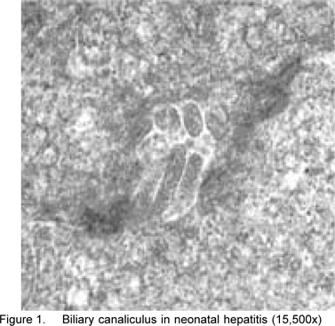
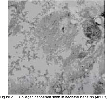
The electron microscopic analysis revealed that in all patients with NH the glycogen content was increased (abundant in 50%), endoplasmic reticulum was dilated and there was increase in mitochondria with abnormal shapes and evidence of degenerative changes (prominent degenerative changes were noted in one patient). Golgi complex was seen in 50% patients with no specific features. Dilated bile canaliculi were seen in only one patient with evidence of epithelial fragments in the lumen suggestive of degenerative changes (Figures 1 and 2). No changes pertaining to fibrosis were seen in any of the patients. Degenerative myelin figures were seen only in one patient .


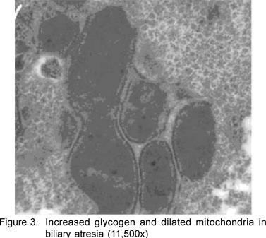
Patients with BA had similar features on electron microscopy. The glycogen content was lowered in one patient while it was abundant in three patients (50%). Endoplasmic reticular changes varied from minimal change in 4 patients to prominent, dilated reticuli in 2 patients. Mitochondrial changes were seen in a majority of patients (66.6%), cytoplasmic degeneration and myelin figures were more prominent in BA (50%). However bile canaliculi seen in one patient did not appear to be dilated, but showed some degenerative cytoplasmic fragments. Collagen deposition corresponding to fibrosis was seen only in one of the two patients with cirrhosis (Figures 3-6).
The light and electron microscopic features of the patients with NH and BA are summarised in Table 2. The electron microscopic features of the two groups of patients were similar with regard to changes in glycogen content and mitochondrial changes. However endoplasmic changes were more pronounced in hepatitis and correlated with ballooning degeneration especially in NH. Cholestatic and cirrhotic changes did not correlate with light and electron microscopic analysis. Also, in one patient with hepatitis, cholestasis was not seen on light microscopy, but only on electron microscopy.

There are very few studies highlighting the comparative light and electron microscopic features of NH and BA and these are also not recent.[5,6,7] Light microscopic changes have been reported before. Park et al[7] found giant cell transformation in 88% and 73%, respectively in the two groups and it was predominantly focal in BA. In our study giant cell transformation was seen in 75% of patients with NH and 33.3% of patients with BA. Intracellular cholestasis was more prevalent (75%) in our study compared to 25% in the study by Park et al in NH. Even in BA, intracellular cholestasis was more prominent than ductular cholestasis (100% vs. 50%). Park et al reported a predominance of ductular cholestasis in BA (82% vs 18%). Ductular proliferation was seen in 50% of NH patients and 100% of BA cases but only to a milder degree in NH. Park et al found ductular proliferation in 91% of BA patients and 13% of NH patients. Extramedullary hematopoiesis and portal inflammation were similar in all patients in both the groups; these were also similar to that reported by Park et al. Fibrosis of portal tracts was seen in only 33.3% in BA compared to 91% in the study by Park et al. Portal fibrosis was also seen in 25% of patients with NH in the study by Park et al, whilst it was not seen in any patient with NH in our study.
From the above it is apparent that the important observation in our study and that in others has been that there is considerable overlap of light microscopic features in both NH and BA. This is significant in assuming that liver biopsy may be diagnostic in differentiating NH from BA.
Electron microscopy revealed prominent endoplasmic changes in all patients with hepatitis, comparable to the study by Park et al (100% vs. 88%). Though endoplasmic changes were more common in BA than those reported (80% vs. 15%), the changes were very mild. Increase in endoplasmic reticulum and dilatation were also reported by Hollander and Schaffner.[5] Degenerative changes in mitochondria were seen in both groups. However, such changes have not been commented on in the study by Park et al but Hollander and Schaffner reported an increase in the number, and irregularity and dilatation of mitochondria. In our study, degenerative cytoplasmic changes in the hepatocytes were more common in NH as compared with BA (100% vs. 50%), whilst Park et al found degeneration was more common in BA (77% vs. 25%). In addition they also noticed that degenerative changes in the biliary ductular epithelium were more prominent in BA (77%). Collagen deposition, signifying inflammatory fibrosis, was seen in one patient with NH and in one with BA. Park et al found fibrosis only in the BA (77%) and not in any of the NH patients. In addition, they also found virus-like particles in two patients with BA, none of which were seen in our study.
Ultrastructural changes in NH were mainly hepatocellular while changes were found to be more pronounced in the periductular region in BA by Park et al. The study by Hollander and Schaffner6 is also significant because they demonstrated relatively few hepatocellular changes in younger patients (7-9 weeks) but more abnormalities in older patients (12-13 months). In our study differences were seen in the hepatocytes between the two groups, mainly in relation to the degenerative endoplasmic reticular changes, cytoplasmic degeneration and occurrence of myelin figures. Ductular changes were similar. Even in the study by Park et al there were patients with NH and BA with indistinguishable ultrastructural features. This lends strong support to Landing’s hypothesis that BA and NH are different manifestations of a single pathological process.[8]
The rarity of the condition, complexity and fixation artifacts during preparation of the grids for electron microscopy have limited the number of patients in this study. Technical limitations have not allowed us to study ductular changes in greater detail; more grids for each patient may have allowed more detailed evaluation of the canaliculi and ductular and portal changes. The small number of patients also limited the evaluation of changes in BA with reference to age at surgery and for specific prognostic features.
However, despite the limitations of the study, the following conclusions can be drawn:
- Ultrastructural changes in neonatal cholestasis as studied by electron microscopy are largely non-specific and serve poorly to differentiate BA from NH, at least in patients older than 60 days.
- Dilated endoplasmic reticulum and mitochondrial degeneration correlate with ballooning degeneration seen on light microscopy and hence are more common in NH.
- Cholestatic changes do not correlate with light and electron microscopic findings in all patients.
Idiopathic hepatitis and biliary atresia probably represent varied manifestations of a similar insult, resulting in similar utlrastructural hepatocyte features. A larger study enrolling patients at various ages of presentation and follow up biopsies in the postoperative period might help to unravel the hiddenmysteries concerning biliary atresia.
References
1. Sokol RJ, Mack C, Narkewicz MR, Karrer FM. Pathogenesis and outcome of biliary atresia: current concepts. J Pediatr Gastroenterol Nutr. 2003;37:4–21.
2. Balistreri WF, Grand R, Hoofnagle JH, Suchy FJ, Ryckman FC, Perlmutter DH, et al. Biliary atresia: current concepts and research directions. Summary of a symposium. Hepatology. 1996;23:1682–92.
3. Kasai M, Mochizuki I, Ohkohchi N,Chiba T, Ohi R. Surgical limitations for biliary atresia; indications for liver transplantation. J Pediatr Surg. 1989;24:851–4.
4. Krishnamurthy GT, Turner FE. Pharmacokinetics and clinical application of technetium 99m-labeled hepatobiliary agents. Semin Nucl Med. 1990;20:130–49.
5. Hollander M, Schaffner F. Electron microscopic studies in biliary atresia. I. Bile ductular proliferation. Am J Dis Child. 1968;116:49–56.
6. Hollander M, Schaffner F. Electron microscopic studies in biliary atresia II. Hepatocellular alterations. Am J Dis Child. 1968;116:57–65.
7. Park WH, Kim SP, Park KK, Choi SO, Lee HJ, Kwon KY. Electron microscopic study of the liver with biliary atresia and neonatal hepatitis. J Pediatr Surg. 1996;31:367–74.
8. Landing BH. Considerations of the pathogenesis of neonatal hepatitis, biliary atresia and choledochal cyst—the concept of infantile obstructive cholangiopathy. Prog Pediatr Surg. 1974;6:113–39.