48uep6bbphidvals|275
48uep6bbphidcol4|ID
48uep6bbph|2000F98CTab_Articles|Fulltext
Up to 90% of gastrointestinal foreign bodies (GIFBs) pass spontaneously through the digestive tract without inflicting harm on the patient.[1] However, there is always the chance of injury. The mortality associated with GIFBs that do not pass spontaneously is as high as 50%. Bowel perforation and obstruction are the most common significant complications associated with GIFBs. Other complications include bleeding, respiratory compromise, fistula formation, and abscess formation. In patients presenting with symptoms related to a GIFB, the perforation rate has been estimated to be as high as 5% (up to 35% for sharp-pointed objects). The objects most frequently ingested by children are coins. Objects that become entrapped in the ileocecal valve tend to be smaller, because they have first to pass through the pylorus, duodenal sweep, and ligament of Treitz. Most objects are passed within 4 to 6 days, although some may take as long as 4 weeks. The convention that we follow is that once the coin passes the stomach, and is lodged in a given location for more than 1 week, surgical removal must be considered. Most reports of small bowel coin impaction have been managed surgically. We report the endoscopic management of a 12-year child who had an impacted coin at the ileocecal valve and developed small bowel obstruction.
Case Report
We report a case of small bowel obstruction secondary to ingestion of a one-rupee coin, which was removed endoscopically.
A 12-year-old boy presented to the Emergency Department (ED) with a one-week history of abdominal pain and vomiting. His history included the accidental ingestion of a one-rupee coin seven days ago following which he developed abdominal pain, distension and vomiting. Radiographic examination with x-ray abdomen showed the radiopaque shadow of the coin in the pelvis overlying the right iliac bone along with multiple air fluid levels in the left upper abdomen. Chest x-ray was normal.
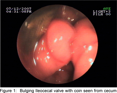
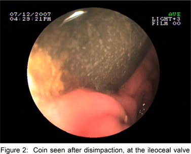
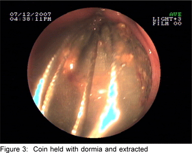
Persistent abdominal complaints, abnormal physical examination, and failure of the coin to progress through thegastrointestinal tract even after seven days despite conservative management led to endoscopic management with colonoscopy. Oral colonoscopy preparation was not attempted in view of bowel obstruction. Therefore colonoscopy was performed under conscious sedation following bowel preparation with enema. Colonoscopy showed bulging and edematous ileocecal valve (Figure 1) and on suction the impacted coin was visible through the ileocecal valve. The colonoscope was used to disimpact the coin (Figure 2) and push it back in the terminal ileum. The colonoscope was negotiated through the ileocecal valve into the terminal ileum. The coin was grasped with a dormia (Figure 3) and pulled through the ileocecal valve and removed per anus. After removal of the coin the patient improved and the abdominal pain and distension remitted. Repeat x-ray abdomen showed that the small bowel air fluid levels had disappeared.
Discussion
There have been reports of cases of small bowel obstruction secondary to coin ingestion, which have been managedsurgically. In one such report, of a 22-year-old woman who had a 3-week history of abdominal pain, surgical exploration revealed that the coin had lodged near the ileocecal valve and an inflammatory mass had formed around the intra-luminal coin, resulting in the formation of a 10 cm x 7 cm fibrous tumour completely obstructing the small bowel. It was thought that oxidation of the coin, with subsequent exposure of its high zinc content, instigated the inflammatory cascade.[2]
In a retrospective study of 104 children who had ingested foreign bodies, the overall success rate for endoscopic management was 98.8%. There were no complications during endoscopic intervention. The authors concluded that foreign bodies in the small and large intestines tended to pass through spontaneously without complications.[3] However, in our case the coin was impacted at the ileocecal valve and led to small bowel obstruction, which required endoscopic removal. After removal of the coin the patient recovered completely.
Conclusion
In children, an impacted foreign body at the ileocecal valve with small bowel obstruction can be removed endoscopically under conscious sedation. Colonoscopic removal of such foreign bodies is an effective and safe method. The method also prevents erosion and perforation of the gastrointestinal tract and relieves the small bowel obstruction. This report suggests that endoscopic removal should be attempted in small bowel obstruction. Now with the availability of modern enteroscopes it may be possible to manage a subset of such small bowel foreign body impactions and obstructions endoscopically.
References
1. Schwartz GF, Polsky HS. Ingested foreign bodies of the gastrointestinal tract. Am Surg 1976;42:236–8.
2. Tupesis JP, Kaminski A, Patel H, Howes D. A penny for your thoughts: small bowel obstruction secondary to coin ingestion. J Emerg Med. 2004;27:249–52.
3. Kim JK, Kim SS, Kim JI, Kim SW, Yang YS, Cho SH et al. Management of foreign bodies in the gastrointestinal tract: an analysis of 104 cases in children. Endoscopy. 1999;31:302–4.