48uep6bbphidvals|258
48uep6bbphidcol4|ID
48uep6bbph|2000F98CTab_Articles|Fulltext
Mucormycosis is a rare opportunistic infection caused by fungi of the order mucorales viz. mucor, rhizomucor, absidia, apophysomyces etc[1]. These infections are frequently encountered in immunocompromised host[2,3], however, its occurrence in immunocompetent host is not unknown [4,5]. To the best of our knowledge, only one case of isolated renal with splenic mucormycosis has been reported in the literature and the patient could not be salvaged[6].
Case Report
A 28-years male was hospitalized in with a history of fever, pain in the left flank and loss of appetite of 2 weeks duration. The patient had no history of diabetes mellitus, use of steroids, trauma or sexual exposure. He had a toxic look and was febrile. His pulse was 124/min and blood pressure 110/ 80mmHg.Abdominal examination revealed tenderness and guarding in the left hypochondrium and left lumbar region. Other systemic examination was normal. Haemogram showed haemoglobin of 10.8g/dl and leukocytosis (21,000/ mm3) with 82% polymorphs. The urine analysis revealed microscopic haematuria and pyuria (20-25 cells/HPF). The serum creatinine was 1.1mg/dl and blood urea 50mg/dl. Blood sugar was normal. Liver function tests revealed mild hyperbilirubinemia (2.1 mg%) and elevated serum alkaline phosphatase (25 KAU/ml: normal 3-16 KAU/ml). Blood and urine cultures for pyogenic organisms and fungi were negative. The viral serology including HIV 1 and 2, HBsAg and HCV were negative.
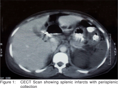
Chest x-ray was normal. The ultrasonography of the abdomen showed a hypoechoic lesion in the lower pole of spleen. The patient did not respond to antibiotics. Subsequent contrast enhanced CT scan performed which showed an enlarged left kidney with multiple infarcts, perinephric inflammatory changes and splenic infarct with perisplenic collection (Figure 1). Smear examination of the ultrasound guided aspirate from perisplenic collection revealed broad irregular aseptate hyphae.
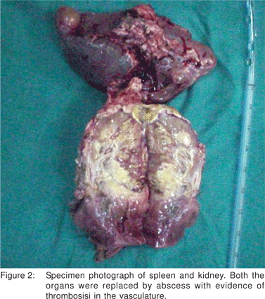
In view of the above, amphotericin B was started and the patient underwent splenectomy, left nephrectomy and debridement. The spleen was replaced by a large cavity containing necrotic material. The splenic artery and vein were thrombosed. Left kidney was infarcted and showed areas of necrosis. The left renal artery and vein were thrombosed (Figure 2).
The histopathology showed massive splenic infarcts with very little preserved splenic tissue and few broad aseptate fungal hyphae with right angle branching suggestive of mucormycosis. The splenic artery was thrombosed with fungal invasion. The kidney revealed multiple fresh infarcts (Figure 3) in both the cortex and the medulla.
The patient received amphotericin B for 60 days at a dose of 50mg/kg/day (total dose of 3gms). Follow up ultrasonography of abdomen at 3 and 6 months showed noresidual collection. At 42 months follow-up the patient is well.

Discussion
Mucorales are ubiquitous saprophytic fungi found in decaying vegetative and organic matter[1,2,6]. The fungi and their spores have minimal intrinsic pathogenicity but they can initiate aggressive and often fatal infection in immunocompromised patients[6,7]. The occurrence of mucormycosis in a previously healthy individual is rare[4,5,6]. However, few cases of mucormycosis in immunocompetent patients have been reported from our center[4,6].
Depending on the portal of entry viz infections through inhalation, ingestion, contamination of skin wounds or via a vascular channel such as an intravenous drip, well known clinical forms of mucormycosis are known to develop[6]. The most common form is rhinocerebral followed by pulmonary, cutaneous, gastrointestinal and disseminated [6,7,8]. In the disseminated form, the most commonly involved organ is the lung followed by brain, kidney, heart, spleen etc[9]. Isolated renal with splenic mucormycosis is extremely rare [3,10]. The mode of infection of the kidney is controversial. Spared ureters and bladder in isolated bilateral renal involvement supports the hematogenous spread[3,10]. Simultaneous involvement of the left kidney and spleen is suggestive of contiguous spread of infection from the left kidney to the spleen or vice-versa through leinorenal ligament. Likewise, the ureters and bladder were not involved in the index case. However, haematogenous mode of infection of both the organs cannot be ruled out.
The characteristic feature of mucor infection is the vascular invasion with thrombosis involving large and small arteries, which results in infarction and necrosis of the affected organ[1,2,4,6,7]. Rarity of this condition leads to misdiagnosis at initial presentation. An early diagnosis requires a high index of clinical suspicion and demonstration of fungal hyphae on smear examination of aspirated fluid[11]. Mucorales are identified by their broad aseptate hyphae branching at right angles at irregular intervals[1,6,7,11].
The mortality rate of mucormycosis is very high and the average survival rate is 36%[8,9,11]. The highest mortality is seen with the disseminated disease and the lowest is seen in infections confined to skin and subcutaneous tissue[8,9,11]. Successful treatment requires an early diagnosis and aggressive surgical debridement of infected tissues, combined with antifungal therapy[1,6,7,8,9,11].
Concluding, mucormycosis although common in immunocompromised patients, can also invade immunocompetent hosts. Isolated splenic with renal involvement in the absence of systemic infection is extremely rare. The possible portal of infection seems to be haematogenous or contiguous spread. High index of clinical suspicion and characteristic CT findings leads to early diagnosis. Successful management lies in the aggressive surgical debridement of infected tissues with antifungal therapy.
References
1. Eucker J, Sezer O, Graf B, Possinger K. Mucormycosis. Mycoses 2001;44:253–60.
2. Turunc T, Demiroglu YZ, Aliskan H, Colakoglu S, Arslan H. Eleven cases of mucormycosis with atypical clinical manifestations in diabetic patients. Diabetes Res Ciln Pract. 2008;82:203–8.
3. Aslani J, Eizadi M, Kardavani B, Khoddami-Vishteh HR, Nemati E, Hoseini SM, et al. Mucormycosis after kidney transplantations: report of seven cases. Scand J Infect Dis. 2007;39:703–6.
4. Sharma A, Gupta V, Singh RS, Kakkar N, Singh S, Bambery P. Angioinvasive pulmonary mucormycosis presenting as multiple bilateral pulmonary nodules in a patient without obvious predisposing factors. Singapore Med J. 2008;49:e269–71.
5. Thomas AJ, Shah S, Mathews MS, Chacko N. Apophysomyces elegans - renal mucormycosis in a healthy host: a case report from south India. Ind J Med Microbiol 2008;26:269–71.
6. ChakrabartiA, Ghosh A, Prasad GS, David JK, Gupta S, Das A, et al. Apophysomyces elegans: an emerging zygomycete in India. J Clin Microbiol 2003;41:783–8.
. Benbow EW, Stoddart RW. Systemic zygomycosis. Postgrad Med J. 1980;62:985–96.
8. Sims CR, Ostrosky-Zeichner L. Contemporary treatment and outcomes of zygomycosis in a non-oncologic tertiary care center. Arch Med Res. 2007;38:90–3.
9. Rinaldi MG. Zygomycosis. Infect Dis Clin North Am. 1989;3:19–41.
10. Singh SK, Wadhwa P, Sakhuja V. Isolated bilateral renal mucormycosis. Urology. 2004;63:979–80.
11. Safar A, Marsan J, Marglani O, Al-Sebeih K, Al-Harbi J, Valvoda M. Early identification of rhinocerebral mucormycosis. J Otolaryngol. 2005;34:166–71.