Original Articles
 
Increased prevalence of type II diabetes mellitus in hepatitis C virus infection in Western India
 
Deepak, N Amarapurkar, Nikhil D Patel
Department of Gastroenterology,
Bombay Hospital and Medical Research Centre,
Mumbai, India


Corresponding Author
:
Dr. Nikhil D. Patel
Email : niyopatel@yahoo.com


Abstract

Background: Type II diabetes mellitus (DM) has been shown as more common in patients with hepatitis C virus infection (HCV). Similar data from India is not available. Methods: This 3-year prospective study included consecutive Indian patients with HCV to detect the DM. In all patients, the presence of DM, duration of DM, probable duration of HCV, genotype of HCV, presence of steatosis and presence of cirrhosis were noted. Comparable numbers of consecutive patients with hepatitis B virus infection (HBV) and irritable bowel syndrome (IBS) were analysed for the presence of DM. Results: A total of 200 patients with HCV were analysed: mean age=45.9±9.8 years; male:female=1.3:1; genotype distribution (in 80 patients which included 17 patients of DM)- genotype 3 in 47(58%), genotype 1 in 31(39%) and genotype 2 in 2(3%) patients; probable duration (unknown in 40 patients) of  HCV=12.8±8.2 years; steatosis in 55(27.5%) patients; cirrhosis in 88(44%) patients. Of these 200 patients, DM was present in 44(22%) patients with mean duration of DM of 6.1±2.3 years. HCV preceded DM in 29 patients by 10.8±2.3 years. Among HCV with genotype 3, DM was present in 11(23.4%) patients and with genotype 1, DM was present in 6(19.3%) patients. In patients with DM, cirrhosis and steatosis were present in 28(63.6%) and 20(45.4%) patients, respectively, as compared to 60(38.4%) and 35(22.4%) patients without DM. There was significantly lower presence of DM, 24(12%) and 19(9.5%), in 200 patients of HBV and 200 patients of IBS, respectively. Conclusion: There is increased prevalence of DM in patients with HCV. HCV precedes the development of DM by a decade.


48uep6bbphidvals|247
48uep6bbphidcol2|ID
48uep6bbph|2000F98CTab_Articles|Fulltext

Hepatitis C virus (HCV) infection is poorly linked to many extrahepatic manifestations except essential mixed cryoglobulinemia and membranous proliferative glomerulonephritis.[1]

Type II diabetes mellitus (DM) is commoner in HCV infected patients (19-62%) than in other control groups (3-13%).[2,3,4,5,6] The risk of developing DM increases 2-3 times in the presence of HCV.[3,6] There was increased incidence of post-(liver/renal) transplantation DM in HCV infected recipients[7] and in recipients who received kidneys from HCV infected donors.[8] Successful interferon treatment improves DM control.[9] Also, HCV infection is higher (4.2-28%) in DM than in the general population (0.5- 2.5%).[10] DM is associated with rapid progression of liver disease and with development of hepatocellular carcinoma in HCV.[10]


However, the causal association of DM and HCV is not clear. It is difficult to establish a clear link in the presence of traditional risk factors for the development of insulin resistance (IR) and thereby DM: age=45 years, male sex, race, obesity, dyslipidemia, hypertension, family history of DM, past history of impaired glucose tolerance, history of delivery of large babies, history of gestational diabetes and polycystic ovary syndrome.[11] DM itself increases the risk of chronic liver disease and even hepatocellular carcinoma.[12] Mortality from cirrhosis is higher in DM than cardiovascular causes.[13] Also, cirrhosis is considered a diabetogenic condition irrespective of the aetiology of cirrhosis.[14]


Contrary to the above studies, few studies have failed to show any relation between DM and HCV and reported low prevalence of DM in HCV[14] and also low prevalence of HCV in DM.[15]


Prevalence of DM in the general population of India has gone up from < 2% before 1977 to > 12% since 2000.[11] In India, the prevalence of HCV ranges from 0.3 to 4% in the general population.[16] There are only a few studies from India to document the relationship between the two diseases in question.[17,18] Most studies in India have shown a predominance of HCV genotype III with prevalence ranging from 12% to 64%.[19] But effect of HCV genotype III on the prevalence of DM is not widely known.


We previously reported a higher incidence of DM in chronic hepatitis C virus infection (CHC) (14%) as compared to chronic hepatitis B virus infection (CHB) (8.5%) and general population (5%), prevalence of DM being the same in patients with cirrhosis in CHC and CHB.[17] In another series when looking at the spectrum of chronic liver disease in diabetics, we found that CHC had similar prevalence, whereas prevalence of CHB was lower in diabetics compared to non-diabetics.[20] In another recent study from North India, the prevalence of DM was similar in the HCV infected (12.5%) and in the general population (12- 14%).[18]


Hence, this prospective study was planned to establish the prevalence of DM in CHC, to evaluate the effect of HCV genotypes and stages of liver disease and to compare the prevalence of DM with age and sex-matched patients of CHB and irritable bowel syndrome (IBS).


Methods

Study population


This prospective case-control study was carried out at our centre from January 2002 to December 2004. All consecutive Indian patients with CHC, who were treatment naïve, were evaluated for the presence of DM. Patients with type I diabetes mellitus or HIV infection were excluded. An equal number of patients of CHB and IBS (without clinical, biochemical or imaging evidence of liver disease) seen during this period were also evaluated for DM using similar criteria. Thus, the study population was divided into three groups: group-1 (patients with CHC), group-2 (patients with CHB) and group-3 (patients with IBS).


Evaluation of patients:

Presence of HCV was confirmed by HCV RNA using PCR studies in all cases from group-1 and HCV genotyping was performed using restriction fragment length polymorphism in confirmed cases of HCV whenever possible.


In all the cases, detailed clinical history was taken as follows: symptoms of liver diseases; use of drugs, tobacco and alcohol; list of medications (insulin, oral hypoglycaemic drugs, anti hypertensive drugs and lipid lowering drugs); list of previous laboratory tests chronologically; date of detection of DM (the date when diagnosis of DM established if serial testing was available); family history of DM; risk factors for HCV (blood transfusion, intravenous drug abuse, sexual contact, nosocomial or occupational exposure and maternal HCV status); and probable date of the event leading to HCV (in case of multiple events, date of first-time anti-HCV positivity if serial testing was available or date of first event was taken as the probable date).


All patients had detailed clinical examination noting blood pressure, hepatomegaly, stigmata of chronic liver disease, stigmata of hyperlipidemia and anthropometric parameters (height, weight, waist circumference, hip circumference, body mass index [BMI]).


All patients after 12-hours overnight-fasting underwent complete blood count, liver profile {including alanine aminotransferase [ALT], aspartate aminotransferase [AST], bilirubin, alkaline phosphatase, gamma glutyltransferase, albumin, globulin and prothrombin time}, lipid profile {including total cholesterol, high-density lipoprotein (HDL) cholesterol, low-density lipoprotein cholesterol, triglyceride, apolipoprotein A1, apolipoprotein B and lipoprotein-a}, plasma glucose levels {including random, fasting and post-prandial and/or 2-hour post-75 g glucose levels} and abdominal ultrasonography (for evidence of fatty liver, shrunken and/or nodular liver and portal hypertension). Liver biopsy was performed in groups 1 and 2 whenever possible to evaluate the presence of cirrhosis and steatosis.

Diagnostic criteria:

1.     CHC was defined as the presence of anti-HCV by third generation enzyme immunoassay and HCV RNA positive by polymerase chain reaction for >6 months.

2.     CHB was defined as the presence of serum HBsAg> 6 months.

3.     IBS was defined according to Rome II diagnostic criteria in absence of a structural or biochemical explanation for the symptoms.

4.     DM was diagnosed on the basis of use of oral hypoglycaemic drugs; fasting plasma glucose=126 mg/dl; 2-hour plasma glucose=200 mg/dl during oral glucose tolerance test; and/or random or 2-hour post-prandialplasma glucose=200 mg/dl.

5.     Cirrhosis was diagnosed on the basis of presence of history of decompensation, stigmata of the chronic liverdisease, shrunken and nodular liver and/or portal hypertension on imaging, oesophageal varices on upper gastrointestinal endoscopy and/or presence of cirrhosis on histology (whenever possible).

6.     Steatosis in a non-alcoholic patient (alcohol consumption< 20 g/day) was defined as the presence of fatty liver on imaging and/or presence of macro-vesicular fatty change (>5%) with/without associated necro-inflammation, fibrosis and hepatocyte degeneration on histology (whenever possible).

7.     Aminotransferase elevation was defined as elevation of = 2 times upper limit of normal in AST and/or ALT on two occasions in 6 months.

8.     Dyslipidemia was diagnosed on the basis of the following criteria: fasting cholesterol=200 mg/dl; fastingtriglyceride=150 mg/dl; fasting HDL-cholesterol<40 mg/dl in men or <50 mg/dl in women; and/or use lipid lowering agents.

9.     Hypertension was defined as systolic blood pressure=140 mmHg; diastolic blood pressure=90 mmHg and/or use of antihypertensive agents.

10.   Obesity was defined as BMI=25 kg/m2; waist circumference³ 90 cm (men) or ³80 cm (women); and/or waist- to-hip ratio=0.9 (men) or =0.85 (women).

11.   Family history of DM was defined as the presence of DM in a first-degree relative.


Statistical analysis:


Statistical analysis was performed using the chi square test and student t test (p<0.05 = statistically significant).


Results

Prevalence of DM was higher in group1 compared to other groups, even with comparable presence of risk factors (Table 1).


Comparison of diabetics in all three groups (Table 2) revealed a lower number of patients with age > 40 years, family history of DM, obesity and dyslipidemia in group 1 compared to groups 3 and 2. There were more patients with  steatosis in group 1 compared to group 2, but presence of cirrhosis was comparable in both groups.


In diabetics from groups 1 and 2, there were more patients with aminotransferase elevation, cirrhosis and steatosis as compared to non-diabetics (Tables 3 and 4).


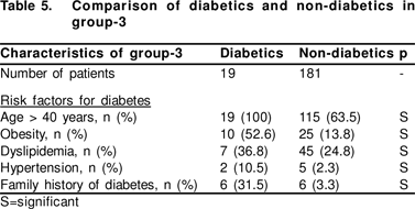
In group 1, diabetics and non-diabetics had comparable risk factors for DM (Table 3). Diabetics of groups 2 and 3 had more patients with risk factors for DM as compared to nondiabetics (Tables 4 and 5). There were no patients with history of delivery of large babies, gestational diabetes or polycystic ovary syndrome in any groups.


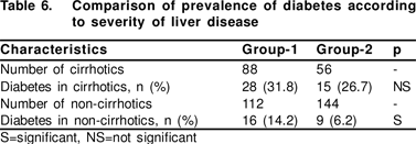
Presence of DM was comparable in cirrhotics of groups 1 and 2, whereas non-cirrhotic patients of group 1 had significantly more DM than group 2 (Table 6).


In group 1, HCV genotype was known in 80 patients, which included 17 patients of diabetes. Genotype III was present in 47 (58%), genotype I in 31 (39%) and genotype II in 2 (3%) patients. There was no difference in genotype distribution in diabetic and non-diabetic patients (Table 3).


Probable date of onset of HCV infection was known in 160 patients from group 1. Of these, 15 patients had history of multiple blood transfusions, 95 patients had history of single blood transfusion and 50 patients were on chronic maintenance haemodialysis. In them, duration of HCV infection was 12.8 ± 8.2 (range=0.5-34) years. In diabetics of group 1, duration of HCV infection was known in 29 patients. In these 29 patients, mean duration of DM was 6.1 ± 2.3 (range=0.65- 30) years. HCV infection preceded DM in 29 patients by 10.8 ± 2.3 (range=0.1-26) years.


Discussion

Our study demonstrated a higher prevalence of DM in CHC, even in non- cirrhotic patients; lower incidence of traditional risk factors in patients with DM and CHC; no relation between prevalence of DM and HCV genotype; and temporal relationship of HCV to DM. These all support a role of HCV in the aetiopathogenesis of DM.


Our study showed increased prevalence of DM in CHC compared to CHB, which is in accordance with previous reports.[4,5]


Our study could not identify patients with increased risk factors for the development of DM in CHC. In previous series, HCV was shown to increase the chances of development of DM in the presence of traditional risk factors: non-white race,[3] genotype 2a or 1a,[4] severity of underlying liver disease or presence of cirrhosis,[1,2,5] increased age,[3,4,5,6] male sex,[6] obesity,[3,6] low socioeconomic class,3 previous interferon treatment,[21] or family history of DM.2 In a recent series, it was shown that diabetics in HCV infection did not show classical phenotypic correlates, but presented at a younger age, and had lower rates of obesity and dyslipidemia.[22] Together with our series, these findings support the concept that DM in CHC represents two types of patients i.e. one related to traditional risk factors and one related to the HCV infection itself.


In our series, there was no effect of HCV genotypes on the development of DM. Most extrahepatic manifestations are not genotype-specific.[1]


Temporal relationship with HCV infection and development of DM is difficult to estimate accurately, as both disorders have insidious onset. Our study showed that in patients where the exact duration of both DM and HCV infection was known, HCV infection preceded the development of DM by a decade, even in the absence of traditional risk factors.


Diabetics in our series had increased prevalence of aminotransferase activity, cirrhosis and steatosis, thereby suggesting a role of DM in the progression of liver disease of any cause. Non-alcoholic steatosis is considered a part of the metabolic syndrome and is closely associated with IR and DM. [23] In our study, diabetics with CHC were more likely to have steatosis than were CHB patients, this was in accordance to previous reports.[24]


In our study, steatosis was seen more often in CHC, even when there was increased prevalence of factors responsible for IR in CHB. This was in accordance with previous series as well.[25]


HCV causes IR either by inducing tumour necrosis factor- _,[26] or by altering insulin-signalling pathways.[27] Also HCV induces intrahepatic fat accumulation (steatosis) consequent either of direct effect on hepatocyte,[28] or of hypobetalipoprotienemia induced by HCV[29] or of indirect effect mediated through IR.[10] Steatosis in turn increases IR further[,30] resulting in a vicious cycle, which leads ultimately to the development of DM.[31] Although high ferritin levels are thought to be the result of HCV and are linked to IR, fibrosis and worsening of diabetes,[26] recent studies have shown high ferritin levels to be due to diabetes itself rather than HCV.[32] In addition, a direct cytopathic effect of HCV on pancreatic islet cells has been recently demonstrated[.33] The role of HCVinduced autoimmunity is unlikely to play a major role in the pathogenesis of DM.[4]


All the cirrhotics in our study had increased prevalence of DM regardless of aetiology (whether CHC or CHB). Ourfindings were in accordance with few series,[14] but were contrary to other previous series where cirrhotics with HCV infection were more likely to be diabetics compared to other aetiologies.5 All cirrhotics are predisposed to IR and thereby DM.


As non-cirrhotics also have increased prevalence of DM in CHC, it is unlikely that DM in HCV infection reflects only the severity of underlying liver disease, as was suggested by a few authors.[4] Our results were in accordance with previous series showing increased DM in non-cirrhotic HCV infection.[3,27,32]


In conclusion, HCV appears to be a predisposing and preceding factor for the development of DM, even in  noncirrhotics and presents typically, i.e. without the presence of traditional risk factors. 


References

1.     Agnello V, De Rosa FG. Extrahepatic disease manifestations of HCV infection- some current issues. J Hepatol. 2004;40:341–52.

2.     Zein CO, Levy C, Basu A, Zein NN. Chronic hepatitis C and type II diabetes mellitus- a prospective cross-sectional study. Am J Gastroenterol. 2005;100:48–55.

3.     Mehta SH, Brancati FL, Sulkowski MS, Strathdee SA, Szklo M, Thomas DL. Prevalence of type 2 diabetes mellitus among persons with hepatitis C virus infection in the United States. Ann Intern Med. 2000;133:592–9.

4.     Mason AL, Lau JY, Hoang N, Qian K, Alexander GJ, Xu L, et al. Association of diabetes mellitus and chronic hepatitis C virus infection. Hepatology. 1999;29:328–33.

5.     Caronia S, Taylor K, Pagliaro L, Carr C, Palazzo U, Petrik J, et al. Further evidence for an association between non- insulin dependent diabetes mellitus and chronic hepatitis C virus infection. Hepatology. 1999;30:1059–63.

6.     Grimbert S, Valensi P, Levy-Marchal C, Perret G, Richardet JP, Raffoux C, et al. High prevalence of diabetes mellitus in patients with chronic hepatitis C- a case-control study. Gastroenterol Clin Biol. 1996;20:544–8.

7.     Mora PF. Post-transplantation diabetes mellitus. Am J Med Sci. 2005;329:86–94.

8.     Abbott KC, Lentine KL, Bucci JR, Agodoa LY, Koff JM, Holtzmuller KC, et al. Impact of diabetes and hepatitis af ter kidney transplantation on patients who are affected by hepatitis C virus. J Am Soc Nephrol. 2004;15:3166–74.

9.     Tanaka H, Shiota G, Kawasaki H. Changes in glucose tolerance after interferon-alpha therapy in patients with chronic hepatitis C. J Med. 1997;28:335–46.

10.   Hui JM, Sud A, Farrell GC, Bandara P, Byth K, Kench JG, et al. Insulin resistance is associated with chronic hepatitis C virus infection and f ibrosis progression. Gastroenterology. 2003;125:1695–704.

11.   Iyer SR. Type 2 diabetes mellitus express highway, where is the ‘U’ turn? J Ass Physc India. 2003;51:495–500.

12.   El-Serag HB, Tran T, Everhart JE. Diabetes increases the risk of chronic liver disease and hepatocellular carcinoma. Gastroenterology. 2004;126:460–8.

13.   de Marco R, Locatelli F, Zoppini G, Verlato G, Bonora E, Muggeo M. Cause-specific mortality in type 2 diabetes- the Verona Diabetes Study. Diabetes Care. 1999;22:756–61.

14.   Mangia A, Schiavone G, Lezzi G, Marmo R, Bruno F, Villani MR, et al. HCV and diabetes mellitus- evidence for a negative association. Am J Gastroenterol. 1998;93:2363–7.

15.   Sotiropoulos A, Peppas TA, Skliros E, Apostolou O, Kotsini V, Pappas SI. Low prevalence of hepatitis C virus infection in Greek diabetic Patients. Diabetes Med. 1999;16:250–2.

16.   Arankalle VA. Epidemiology of HCV infection in India: a comprehensive analysis. In Hepatitis B and C- carrier to cancer. Sarin SK, Okuda K eds. Haurcourt India Pvt Ltd. 2002;201–18.

17.   Amarapurkar DN. Is diabetes mellitus an extrahepatic manifestationof hepatitis C infection? Indian J Gastroenterol. 1999;18:A43.

18.   Duseja A, Sharma S, Nitin, Chaudhry S, Dhiman RK, Chawla Y, et al. Prevalence of diabetes mellitus in patients with chronic hepatitis C virus infection. J Gastroenterol Hepatol. 2004;19:A711–2.

19.   Amarapurkar D, Dhorda M, Kirpalani A, Amarapurkar A, Kankonkar S. Prevalence of hepatitis C genotypes in Indian Patients and their clinical significance. J Ass Physc India. 2001;49:983–5.

20.   Amarapurkar D, Das HS. Chronic liver disease in diabetes mellitus. Tropical Gastroenterol. 2002;23:3–5.

21.   Imano E, Kanda T, Ishigami Y, Kubota M, Ikeda M, Matsuhisa M, et al. Interferon induces insulin resistance in patients with chronic active hepatitis C. J Hepatol. 1998;28:189–93.

22.   Behrendt CE, Ruiz RB. Hyperglycemia among persons with hepatitis C- not the classical diabetic phenotype. Diabetes Research Clinc Practice. 2006;71:68–74.

23.   Amarapurkar DN, Patel ND. Clinical spectrum and natural history of non-alcoholic steatohepatitis with normal alanine aminotransferase values. Tropical Gastroenterology. 2004;25:130-4.

24.   Czaja AJ, Carpenter HA, Santrach PJ, Moore SB. Host and disease specific factors affecting steatosis in chronic hepatitis C. J Hepatol. 1998;29:198–206.

25.   Sanyal AJ, Contos MJ, Sterling RK, Luketic VA, Shiffman ML, Stravitz ML, et al. Nonalcoholic fatty liver disease in patients with hepatitis C is associated with features of the metabolic syndrome. Am J Gastroenterol. 2003;98:2064–71.

26.   Elsammak M, Refai W, Elsawaf A, Abdel-Fattah I, Abd Elatti E, Ghazal A. Elevated serum tumour necrosis factor alpha and ferritin may contribute to the insulin resistance found in HCV positive Egyptian patients. Cur rent Med Research Opinion. 2005;21:527–34.

27.   Kawaguchi T, Yoshida T, Harada M, Hisamoto T, Nagao Y, Ide T, et al. hepatitis C virus down-regulates insulin receptor substrates 1 and 2 through up-regulation of suppressor of cytokine signalling 3. Am J Pathol 2004;165:1499–508.

28.   Lerat H, Honda M, Beard MR, Loesch K, Sun J, Yang Y, et al. Steatosis and liver cancer in transgenic mice expressing the st ructural and nonstructural proteins of hepatitis C virus. Gastroenterology. 2002;122:352–65.

29.   Serfaty L, Andreani T, Giral P, Carbonell N, Chazouilleres O, Poupon R. Hepatitis C virus induced hypobetalipoproteinemia- a possible mechanism for steatosis in chronic hepatitis C. J Hepatol. 2001;34:428–34.

30.   Chou CJ, Haluzik M, Gregory C, Dietz KR, Vinson C, Gavrilova O, et al. WY14,643, a peroxisome proliferatoractivated receptor alpha (PPARalpha) agonist, improves hepatic and muscle steatosis and reverses insulin resistance in lipoatrophic A-ZIP/F- 1 mice. J Biological Chem. 2002;277:24484–9.

31.   Noto H, Raskin P. Hepatitis C infection and diabetes. J Diabetes Complications. 2006;20:113–20.

32.   Lecube A, Hernandez C, Genesca J, Esteban JI, Jardi R, Garcia L, et al. Diabetes is the main factor accounting for the high ferritin levels detected in chronic hepatitis C virus infection. Diabetes Care. 2004;27:2669–75.

33.   Masini M, Campani D, Boggi U, Menicagli M, Funel N, Pollera M, et al. Hepatitis C virus infection and human pancreatic beta-cell dysfunction. Diabetes Care. 2005;28:940–1.