Vivek Agrawal, Naveen Sharma, Mohit Kumar Joshi, VR Minocha
Department of Surgery
University College of Medical Sciences and Guru Teg Bahadur Hospital
Delhi, India
Corresponding Author:
Dr. Vivek Agrawal
Email: vivekgtbh @gmail.com
Abstract
Background: There are no accepted guidelines for the closure of laparotomy incisions inpatients of peritonitis. As these patients differ from the patients undergoing electiveabdominal surgery, the same recommendations for closure may not be applicable in bothgroups.
Aim: To compare wound outcome parameters following closure of the laparotomy incisionwith absorbable and non-absorbable suture material using the continuous and interruptedtechniques in patients of peritonitis.
Method: A single blinded randomised controlled trial using Polygalactin–910 andPolypropylene, number 1 sutures, to close midline vertical incisions, placed in continuousand interrupted manner, was performed on 174 patients. Patients were randomised intofour groups: Group A (Polygalactin-910 continuous suturing, n=40), B (Polygalactin-910interrupted suturing, n=47), C (Polypropylene continuous suturing, n=45) and D(Polypropylene interrupted suturing, n=42). The incidence of wound infection, dehiscence,suture sinus formation and incisional hernia was recorded. Patients were followed up fora period of four years. Statistical analysis involved the chi-square and Fisher’s exact tests.A ‘p’ value of <0.05 was considered significant.
Results: The study included 139 male and 35 female patients between the ages of 10 and75 years. The incidence of wound infection (p=0.656), dehiscence (p=0.997), and incisionalhernia (p=0.930) at 3 months and four years (p= 0.910) was not statistically significant.There was no sinus formation in groups A and B, however 2 patients of group C and 6patients of group D did develop suture sinus (p=0.003).
Conclusion: Suture material and technique of closure does not influence wound outcomein patients of peritonitis except for a significantly lower incidence of sinus formation whennon-absorbable sutures are used.
|
48uep6bbphidvals|181 48uep6bbphidcol4|ID 48uep6bbph|2000F98CTab_Articles|Fulltext There are no guidelines in the principles of technique andtype of suture material for incision closure in patientsundergoing emergency abdominal surgery for peritonitis. Nostudy dealing in wound performance in patients of peritonitiswas found in a Medline search except for a retrospectivestudy on the incidence of incisional hernia.[1] The recommendations for abdominal wall closure in patientsundergoing elective abdominal surgery, have been therefore,extrapolated to the patients undergoing emergencyabdominal surgery. Candidates undergoing emergencyabdominal surgery are unwell, poorly optimised, and mayhave peritoneal contamination with risk of intra-abdominalhypertension- factors which may lead to increased incidenceof dehiscence and wound infection. Thus, such a subgroupof patients cannot be compared to the patients undergoingelective abdominal surgery. The rate of wound complications like dehiscence and hernia after laparotomy has remainedfairly consistent during the last 75 years[2] despite the availability of newer and better types of suture material andincorporation of the recommendations for closure of theabdominal wound[3] into routine surgical practice. The use ofnon-absorbable suture material has been supported byseveral studies with the premise that the wound neverachieves the original strength as that of prior to surgery.[4] Ithas been reported that six weeks after incision, the fascialwound achieves only 60%-80% of its pre-operative strength,[5] leading to the belief that sutures are unimportant in adding tothe healing of an abdominal wound beyond this period.However, manufacturers often promote one suture over theother, quoting small series of supposedly superior results.This study attempts to clarify whether suture material(absorbable or non-absorbable) and technique of closure (interrupted or continuous) affect the wound outcome inpatients of peritonitis undergoing emergency abdominalsurgery.
Method
All peritonitis cases of either sex, presenting to the UniversityCollege of Medical Sciences and Guru Teg Bahadur Hospital,a tertiary care teaching hospital of Delhi, India, from the 1st of March 2000 to the 30th of April 2002 were assessed. The study was approved by the institutional ethics committee.APACHE II score and intra-abdominal pressure (by measuringintravesical pressure) were calculated for all patients, inaddition to routine hematological and biochemical tests.Informed consent was taken from all patients for surgery, tobe included in the study. Injection ceftriaxone andmetronidazole were initiated in prescribed pharmacologicaldoses pre-operatively and continued for a period of 7 days.Standard hand washing technique by soap and water wasfollowed by all the members of the operating team. Thesurgical site was painted with 7.5% povidone-iodine solution.Laparotomy was performed by a trained surgeon with aminimum experience of three years of surgical residency. Amiddle midline vertical incision was used, extended aboveor below depending on the location of the pathology and theadequacy of exposure and the relevant operative procedurewas carried out. The patients were randomised by a draw oflots (performed by the staff nurse on floor duty) immediatelyprior to the closure of the abdominal incision, into one of fourgroups: Polygalactin-910 continuous (Group A), Polygalactin-910 interrupted (Group B), Polypropylene continuous (GroupC) and Polypropylene interrupted (Group D). Sutures of sizenumber 1 were employed throughout the trial. Mass closureof the incision was performed in all cases, ensuring adistance of one centimetre (cm) on each side of the marginof the incision and one cm between two adjacent sutures,with suture to wound ratio of 4:1. Anterior and posterior rectussheaths were grasped and drawn medially. The needle waspassed through these layers at the desired distance, asdescribed above, such that the convexity of the needledisplaced the belly of the rectus abdominis muscle laterally.The tension on the suture was just enough to approximatethe margins of the incision. In the interrupted mass closuretechnique each suture was tied separately, and in thecontinuous mass closure technique the suture was tied atthe end of the incision by four throws. The knot was buriedbeneath the repaired rectus sheath in all the patients. Theskin incision was closed using Nylon number 2-0 suture andcovered by an occlusive dressing. The wound was evaluateddaily in the first week for infection and dehiscence, weekly forthe first month for delayed or persistent infection anddehiscence and, fortnightly till three months for sinus formation and incisional hernia. Later, follow-up was carriedout at six monthly intervals for four years to observe if anincisional hernia formed. Once incisional hernia was ruledout on clinical examination, ultrasound examination of theabdomen was done to evaluate the integrity of the scar andpresence or absence of radiologically demonstrable coughimpulse at 3 months, 2 years and 4 years post-operatively todetect any subclinical ventral hernia. Data were collected ona proforma and observations were compiled by an observerblinded, both to the suture used and the technique of closure.
Results
Of the 189 patients recruited in the study, 15 deaths occurredin the early post–operative period (within two weeks) andwere hence excluded from the final analysis. Thus, 174patients were included comprising 139 males and 35 femalecases between the ages of 10 and 75 years. Of the fifteenwho died, 3 were from group A, 6 from group B, 2 from groupC, and 4 from Group D. A total of 11 out of 15 patients expiredwithin the first three days of presentation (having a meanAPACHE II score of 17). Four deaths occurred between Days6 and 14 postoperative (mean APACHE II score 13). Fivedeaths occurred in patients who underwent closure usingthe continuous technique (A & C) and ten in those whounderwent closure using the interrupted technique (B & D).
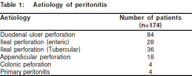
Duodenal ulcer perforation was the most common causeof peritonitis (n= 84, 48.27%) followed by ileal perforation(n=64, 36.78%) (Table 1). The mean APACHE II score of thepatients available for analysis at the end of three months(n=174) was 7 (range 5-14). The intra-abdominal pressurewas comparable amongst the patients of the four groups(mean 18 cm of water). Mean operative time was 68 minutes(35 to 110 minutes). The mean time to suture the incisionwas 11.62 minutes in continuous as compared to 19.92minutes in interrupted suturing. The mean length of suturematerial required for closure was 78 cm in the continuousand 126 cm in the interrupted closure technique. The meanpost-operative stay was 9 days. There was no statisticallysignificant difference in the incidence of wound infection(p=0.656), dehiscence (p=0.997) or incisional hernia at 3months (p=0.930) in between the four groups (Table 2).
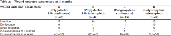
Twenty-three patients were lost to follow up after three monthsleaving 151 patients to be analysed at four years. At 4 years offollow up, although 12 more patients did develop incisionalhernia, the difference was not statistically significant amongstthe four groups (p=0.9). Sinus formation was not seen ingroups A and B (Polygalactin-910), however 2 patients ofgroup C (polypropylene continuous suturing) and 6 patientsof group D (polypropylene interrupted suturing) developedsuture sinus (Table 2); this was statistically significant(p=0.003).
Discussion
Wound related complications of the laparotomy incision arehematoma, infection, dehiscence, suture sinus formation andincisional hernia. The technique of closure and type of suturematerial has been observed to influence wound outcome.[6,7,8,9] Other factors that appear to interfere with wound healing,and can lead to wound failure include advanced age,[9] malnutrition,[9,10,11] hematoma formation, wound infection,[3,9] sepsis,[11] anemia,[9,12] uremia,[12,13] jaundice,[9,12] underlyingmalignancy and corticosteroid therapy.[9,12] Obesity, peritonealdialysis, pulmonary complications, retching, pregnancy andascites, all of which increase the strain on the wound duringthe postoperative period, also predispose to wound healingfailure.[9] There are two types of wound healing failure: early (wounddehiscence) and late (incisional hernia). Rupture of all layersof the abdominal wall with extrusion of the viscera is termedevisceration (burst abdomen). Evisceration occurs inapproximately 1% of laparotomy wounds and is associatedwith a mortality of 15-45%. Infection is associated with almostevery wound that ruptures.[14] Hernia formation is a relativelycommon complication of the abdominal wound with anincidence of 11%, which rises steeply to 23% in the presenceof wound infection.[15] The strength of the abdominal wound lies in themusculoaponeurotic layer. Lichtenstein et al[16] reported thatthe presence of fascial sutures contribute to the strength ofthe abdominal wall for at least 2 months, and that at the timethis has 70% the strength of the unwounded fascia.Dehiscence has been observed with every suture material,yet unfortunately no review of wound dehiscence hasdemonstrated any association between wound disruptionand the use of any other suture material other than catgut. Leaper et al[17] reported that the incidence of wound failurewas 18% for chromic catgut versus 7.8% for polyglycolic acid,monofilament nylon or stainless steel sutures. Cameron etal[18] and Irvin et al[19] were unable to demonstrate any differencein the rates of abdominal wound infection, dehiscence orhernia formation when comparing wounds randomised toclosure with polyglycolic acid or polypropylene sutures.Wissing et al[20] compared 4 methods of closure – continuousand interrupted polygalactin 910, continuous PDS and nylonand found no significant difference. In a meta-analysis ofrandomised clinical trials to determine the influence of typeof suture material and technique of closure on woundcomplications by Hodgson et al,[21] it was found that theincidence of incisional hernia was significantly lower whennon-absorbable sutures were used. This meta-analysis wasin contrast to the findings of our study, wherein we observedno statistically significant difference in the incidence ofincisional hernia in any of the groups at three months and four years of follow up. However, other findings of this meta-analysis including no influence of suture material or techniqueon incidence of wound infection and dehiscence were similaras in our study. The most significant finding in our study wasthe absence of suture sinus formation in patients whounderwent closure with Polygalactin-910 as compared to the8 patients who underwent closure with Polypropylene. Thiswas consistent with the findings of Hodgson et al[21] andGislason et al.[22] Rubio[7] and Gislason et al[22] in their study have concluded that the continuous technique is quicker,cheaper and is as safe as the interrupted technique. Wealso agree with them based on the findings of our study. Ourresults show that the suture materials used and techniquesof closure do not influence any of the wound outcomeparameters, viz., wound infection, dehiscence and incisionalhernia significantly, except for suture sinus formation whennon-absorbable sutures are used.
The number of deaths in the continuous suturing groupsin our study is half that in the interrupted suturing group;though seemingly remarkable this was not statisticallysignificant (p=0.24). Moreover the technique of wound closuredoes not influence the mortality. In cases of peritonitis otherfactors like the patient’s general condition, associated co-morbidities, primary disease process, infection and theduration of illness may also influence wound performance.
Conclusion
Continuous closure of the abdominal incision is preferablein cases of peritonitis because of the economy in time, suturematerial and surgical effort. Interrupted sutures require moretime to apply, and do not confer or impart any additional benefit.The use of absorbable sutures reduces the risk of sinusformation significantly. The incidence of other woundcomplications like infection, wound dehiscence andincisional hernia is not affected by suture material andtechnique of closure, when used in patients being operatedon for peritonitis.
References
-
Mingoli A, Puggioni A, Sgarzini G, Luciani G, Corzani F, CiccaroneF et al. Incidence of incisional hernia following emergencyabdominal surgery. Ital J Gastroenterol Hepatol. 1999;31:449–53.
-
Franz MG, Kuhn MA, Nguyen K, Wang X, Ko F, Wright TE, et al.Transforming growth factor beta (2) lowers the incidence ofincisional hernias. J Surg Res. 2001;97:109–16.
-
Israelsson LA, Jonsson T. Incisional hernia after midlinelaparotomy: A prospective study. Eur J Surg. 1996;162:125–9.
-
Enoch S, Leaper DJ. Basic science of wound healing. Surgery2008;26:31–7.
-
Franz MG. Complications of abdominal wall and hernia operations.In: Mulholland MW, Doherty GM, editors. Complications in surgery. 1st ed. NRC press; 2005;523–45.
-
Israelsson LA, Jonsson T. Closure of midline laparotomy incisionswith polydioxanone and nylon: the importance of suture technique. Br J Surg. 1994;81:1606–8.
-
Rubio PA. Closure of abdominal wounds with continuousnonabsorbable sutures: experience in 1697 cases. Int Surg.1991;76:159–60.
-
Weiland DE, Bay RC, Del Sordi S. Choosing the best abdominalclosure by meta-analysis. Am J Surg. 1998;176:666–70.
-
Begum B, Zaman R, Ahmed M, Ali S. Burst abdomen-A preventablemorbidity. Mymensingh Med J. 2008;17:63–6.
-
Winkler AA, Milburn ML, Holton LT, Goldberg NH, Silverman RP.Effect of suture material on tensile strength and complicationrate in abdominal fascial defects repaired with acellular dermalmatrix. Hernia. 2008;12:33–8.
-
Riou JP, Cohen JR, Johnson H Jr. Factors influencing wounddehiscence. Am J Surg. 1993;166:82.
-
Khan MN, Naqvi AH, Irshad K, Chaudhary AR. Frequency andrisk factor of abdominal wound dehiscence. J Coll PhysiciansSurg Pak. 2004;14:355–7.
-
Makela JT, Kiviniemi H, Juvonen T, Laitinen S.. Factors influencingwound dehiscence after midline laparotomy. Am J Surg.1995;170:387–90.
-
Fleischer GM, Rennert A, Rühmer M. Infected abdominal wall andburst abdomen. Chirurg. 2000;71:754–62.
-
Shukla VK, Mongha R, Gupta N, Chauhan VS, Puneet. Incisionalhernia- comparison of mesh repair with Cardiff repair- a universityhospital experience. Hernia. 2005;9:238–41.
-
Lichtenstein IL, Herzikoff S, Shore JM. The dynamics of woundhealing. Surg Gynecol Obstet. 1970;130:685–90.
-
Leaper DJ, Rosenberg IL, Evans M. The influence of suture materialon abdominal wound healing assessed by controlled trials. EurSurg Res 1976;8:75–6.
-
Cameron AE, Gray RC, Talbot RW, Wyatt AP. Abdominal woundclosure: A trial of Polypropylene and Dexon. Br J Surg.1980;67:487–8.
-
Irvin TT, Koffman CG, Duthie HL. Layer closure of laparotomywounds with absorbable and non- absorbable sutures. Br JSurg. 1976;63:793–6.
-
Wissing J, Van Vroonhoven TJ, Schattenkerk ME, Veen HF,Ponsen RJ, Jeekel J. Fascia closure after midline laparotomy:results of a randomized trial. Br J Surg. 1987;74:738–41.
-
Hodgson NCF, Malthaner RA, Ostbye T. The search for an idealmethod of abdominal fascial closure – a meta analysis. Ann Surg2000;231:436–42.
-
Gislason H, Grønbech JE, Søreide O. Burst abdomen and incisionalhernia after major gastrointestinal operations – comparison ofthree closure techniques. Eur J Surg. 1995;161:349–54.
|