48uep6bbphidcol2|ID
48uep6bbphidvals|1738
48uep6bbph|2000F98CTab_Articles|Fulltext
Esophagitis Dissecans Superficialis (EDS) or sloughing esophagitis is a rare benign illness. The literature on this subject is limited to only a few case reports and case series. EDS is the term given to a rare finding on endoscopy, characterized by the sloughing of esophageal mucosa.
1 The etiology of this condition is unknown and its histopathological features are inadequately described.
Case Report
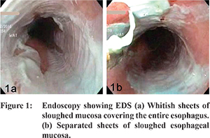
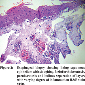
A 54-year-old man presented with history of epigastric discomfort, post-meal fullness and weight loss for the past one month. His medical history was significant for hypertension, hypertriglyceridemia, diabetes mellitus and severe stress. He did not have any history of smoking or consumption of alcohol. He denied any history of nausea, vomiting, gastrointestinal bleeding, dysphagia or odynophagia. There was no history reported of any skin disorders or the use of non-steroidal anti-inflammatory drugs (NSAIDs), bisphosphonates, chemical irritants or hot beverages. His medications included atorvastatin with clopidogrel, losartan, bisoprolol, metformin, glimepiride and multivitamins. On physical examination his vital signs were normal without any organomegaly or palpable abdominal lumps. His hemoglobin was 12g/dl, fasting blood sugar was 109 mg/dl and lipid profile was abnormal. The tests for HIV were negative and serum levels for all tumor markers were normal. Abdominal sonography and CT did not reveal any abnormality. Upper gastrointestinal endoscopy showed white plaque-like lesions affecting the entire esophagus (Figure 1a) with sloughed out mucosa at places (Figure 1b). Biopsy from the lesions demonstrated parakeratosis, orthokeratosis and sloughing of superficial epithelium (Figure 2). A diagnosis of esophagitis dissecans superficialis was confirmed based on these findings. The patients was managed conservatively and follow-up endoscopy and biopsy done after 6 weeks were normal.
Discussion
EDS or sloughing esophagitis is a term applied to the endoscopic finding characterized by sloughing of large fragments of esophageal mucosa that may be coughed up or vomited out as a cast.
1 Although an association with medications (bisphosphonates, potassium chloride, NSAIDs etc.), heavy smoking, hot beverages and autoimmune skin disorders have been reported, most cases are idiopathic.
2-4 While the exact pathogenesis is not known, there may be a common reaction of esophageal squamous mucosa to various types of physical, chemical, thermal, immunological or ischemic insults.
5 Symptoms of EDS can be non-specific or frequently absent. Dysphagia, gastrointestinal bleeding, abdominal pain, heartburn and odynophagia have all been reported in decreasing order of frequency.
6 Endoscopic features of EDS can be patchy or diffuse in the form of white plaques of stripped-off mucosa, which may look like an opaque or translucent membrane, with or without bleeding, vertical fissures and circumferential cracks. The location of the lesions, as reported, was 42% in the distal esophagus, 23% in the entire esophagus, 19% in the middle esophagus and 8% in the proximal esophagus.
6 Biopsy from the confirmed cases shows prominent parakeratosis, orthokeratosis, desquamation of the squamous layers and variable degree of acute or chronic inflammation. There may be an eosinophilic superficial zone with pyknotic or necrotic nuclei and a normal looking basal zone giving rise to a two-toned appearance.
6 There is separation of layers where the basal layers appear viable while the superficial ones slough off and look mummified. In spite of its dramatic presentation at times, EDS resolves spontaneously and completely. A combination of acid suppression and discontinuation of any precipitating drug have been reported to heal the lesions. EDS is a benign condition with an excellent prognosis, which resolves without lasting esophageal pathology because of the superficial nature of the lesions.
References
- Carmack S, Vemulapalli R, Spechler S, et al. Esophagitis Dissecans Superficialis (Sloughing esophagitis): A clinicopathological study of 12 cases. Am J of Surg Pathol. 2009;33:1789-94.
- Gastro Pugh JL, Moore RJ, Wilcox CM, et al. Sloughing esophagitis: a newly recognized clinicopathologic entity. Gastroenterol. 2005;128(Suppl 2):A636-7.
- Cameron RB. Esophagitis dissecans superficialis and alendronate: case report. Gastrointest Endosc. 1997;46:562-3.
- Devarbhavi H, Alvares JF. Esophagitis dessicans superficialis with bulla in chronic renal failure: a case report. Gastrointest Endosc. 2001;54:256-8.
- Cardoso FP, Pinto-Marques P, Neta J, et al. Esophagitis dissecans superficialis. Port Gastrenterol. 2014;21:123-4.
- Purdy J, Appelman H, McKenna B. Sloughing esophagitis is associated with chronic debilitation and medications that injure the esophagus mucosa. Modern Pathology. 2012;25:767-75.