48uep6bbphidcol2|ID
48uep6bbphidvals|1720
48uep6bbph|2000F98CTab_Articles|Fulltext
NAFLD comprises a wide spectrum of liver damage, ranging from simple microvesicular steatosis to steatohepatitis, advanced fibrosis and cirrhosis.
1 In 1980, Ludwig and his colleagues originally coined the term non-alcoholic steatohepatitis (NASH) to describe the morphologic pattern of liver injury in 20 patients evaluated at Mayo clinic over a 10 year period.
2 The term NASH is now reserved to describe progressive forms of NAFLD with degenerative changes and fibrosis.
3 We present the cases of 2 non-obese children below 20 months of age with NASH.
Case History
Case 1
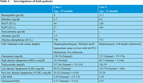
A 19 months old girl born of third degree consanguineous marriage presented with progressive abdominal distension and jaundice for 2 months. There was no bleeding from any site, fever or altered sensorium. Birth history and milestones were normal. There was no illness in other family members. On examination, weight was 7 kg, length was 75 cms. Vital parameters were normal. She had no jaundice. On systemic examination, she had hepatosplenomegaly. Other systems were normal. Investigations are depicted in Table 1. Autoimmune markers (ANF, ASMA, Anti LKM-1) and viral markers ( HBsAg, anti HCV, HIV) were negative. Liver biopsy showed microvesicular and macronodular steatohepatitis with micronodular cirrhosis suggestive of NASH. Her lipid profile is also depicted in Table 1 which showed increased triglycerides and increased very low density lipoproteins (VLDL). Both LDL/HDL ratio and cholesterol/HDL ratio were elevated. Child was advised fat restricted diet and parents were also advised lipid profile. Child is on regular follow up.
Case 2
A 1 year old girl born of third degree consanguineous marriage presented with abdominal distension since 1½ months. There is no jaundice, bleeding or fever. Birth history is normal. There was motor delay in development and child was not able to sit on her own though bisyllable speech was present. On examination, weight was 9 kg, height was 70 cm. She had firm large hepatomegaly. Other systems were normal. Investigations are depicted in Table 1. She was suspected to have a storage disorder and liver biopsy showed micro and macrovesicular fatty liver with fibrosis suggestive of NASH. Her lipid profile is depicted in Table 1 which showed increased cholesterol, triglycerides and LDL Cholesterol. Her serum lactate was 3 mmol/L (Normal = 1-2.4 mmol/L). She was advised fat restricted diet.
Discussion
With the growing prevalence of childhood obesity, pediatric NAFLD has become the most frequent chronic liver disease in children and adolescents in industrialized countries. 9.6% of the American population aged 2 to 19 years has NAFLD, with prevalence rising to 38% in the ones who were obese.
4 It has been suggested that in practice, children with persistently (> 1 year) abnormal liver function tests (> 1.5 times the upper limit of normal) should undergo a systematic assessment to exclude other forms of chronic liver disease and to confirm the diagnosis of NAFLD.
5 Other features such as hypercholesterolemia, and high LDL - cholesterol were also commonly found
6 as seen in our patients where one patient had high cholesterol and the other had high triglycerides. However, they had atypical features, including age of presentation, consanguinity, developmental delay, with a normal weight for age.
Pediatric NAFLD has a distinct histopathological pattern of disease, which is characterized mainly by presence of predominantly portal based injury. It is referred to as type 2 or pediatric NASH to differentiate it from typical zone 3 pattern of disease, which is known as type 1 NASH. However, it was seen that a vast majority of patients had overlapping features of both type 1 adult NASH and type 2 pediatric NASH.
7 Other parameters such as measuring specific circulating markers of fibrogenesis or fibrosis, or changes in tissue elasticity appear to be more promising techniques for staging disease in pediatric NAFLD.
8,9 In a follow up study of non-alcoholic fatty liver disease in children for upto 20 years, Feldstein et al concluded that NAFLD is a disease of progressive potential in children with a significantly shorter long term survival as compared to the general population of the same age and sex. NAFLD in children may progress to cirrhosis and end stage liver disease with the consequent need for liver transplantation, but NAFLD with severe NASH may recur in the allograft.
6 In conclusion, NASH needs to be suspected in cases of children with persistently elevated aminotransferases and further studies need to be conducted in India assess the prevalence of NASH.
References
- Sass DA, Chang P, Chopra KB. Non alcoholic fatty liver disease: A clinical review. Digestive Diseases and Sciences. 2005;50:171-180.
- Ludwig J, Viggiano TR, McGill DB, Oh BJ. Non alcoholic steatohepatitis: Mayo Clinic proc. 1980;55:434-438.
- Baumann U. Brown R. Non alcoholic fatty liver disease in childhood. Br J Diabetes Vasc Dis. 2006;6:264-268.
- Schwimmer JB, Deutsch R, Kahen T, Lavine JE, Stanley C, Behling C. Prevalence of fatty liver in children and adolescents. Pediatrics. 2006;118:1388-1393.
- Angulo P. Non alcoholic fatty liver disease. N Engl J Med. 2002;346:1221-1231.
- Feldstein A E, Charatcharoenwitthaya P, Traprasertsuk S, Benson JT, Enders FB, Angulo P. The natural history of non- alcoholic fatty liver disease in children: a follow up studyfor upto 20 years. Gut. 2009;58:1538-1544.
- Schwimmer JB, Behling C, Newbury R, Deutsch R, Nievergelt C, Schory NJ. Histopathology of pediatric non alcoholic fatty liver disease. Hepatology. 2005;42:641- 649.
- Nobili V, Parkes J, Bottazzo G et al. Performance of ELF serum markers in predicting fibrosis stage in Pediatric Non Alcoholic Fatty Liver Disease. Gastroenterology. 2009;136:160-167.
- Nobili V, Vizzutti F, Alena U, Abraldes JG, Marra F, Pietrobattista A et al. Accuracy and reproducibility of transient elastography for the diagnosis of fibrosis in pediatric non alcoholic steatohepatitis. Hepatology. 2008;48:442-448.