48uep6bbphidvals|708
48uep6bbph|2000F98CTab_Articles|Fulltext
Pancreatic neoplasms (either endocrine or exocrine) occur rarely in children, causing less than 0.2% of malignant pediatric deaths.[1] We report a 2-year-old boy with a multifocal unresectable pancreatoblastoma and a secondary pseudocyst in the tail. This case highlights the unusual presentation of this rare disease, and its diagnostic and management challenges.
Case report
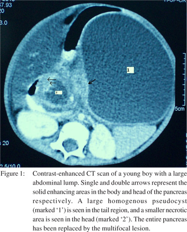
A 2-year-old boy was brought to the surgery clinic with progressive painless left upper abdominal distension of 3 months’ duration. There was no history of pain, vomiting, fever, or, jaundice. General physical examination was unremarkable. On abdominal examination, an 8 cm x 10 cm cystic, non-tender, retroperitoneal swelling was palpable in the left hypochondrium and lumbar region. Ultrasound abdomen was suggestive of a large homogenous hypoechoic lesion in relation to the pancreatic tail, with a bulky pancreatic head. CT abdomen revealed a large, (maximum diameter ~15 cm) cystic, homogenously hypodense lesion in the lesser sac arising from the pancreatic tail, with asymmetric contrast enhancement of the medial wall (Figure 1). The pancreatic body and tail were enlarged, and, two more cystic lesions with multiple solid enhancing areas were identified within the head and body. Fat planes with the superior mesenteric vessels were maintained. A radiological diagnosis of multifocal pancreatoblastoma with a secondary pseudocyst was made. Serum alpha fetoprotein levels were raised. Image-guided FNAC from the mass was inconclusive. Total pancreatectomy was planned. On the day of surgery, the child had fever, tachycardia, and tenderness over the lump. At laparotomy, we found a large, infected, cystic lesion in the lesser sac, densely adherent and inseparable from the posterior gastric wall. Due to dense adhesions and poor general condition, curative resection was deferred. External drainage of the infected pseudocyst was performed, with biopsy of the pancreatic head and a thickened area of the of the cyst wall. The patient recovered satisfactorily in the postoperative period.
Histopathological examination revealed small round epitheloid cells with atypical nuclei arranged in uniform nests. A final diagnosis of multifocal pancreatoblastoma with a secondary pseudocyst was made, and the patient was planned for chemotherapy.
Discussion
Pancreatoblastoma and solid-papillary epithelial tumor are the most common pancreatic neoplasms in children. Only about 200 cases of pancreatoblastoma have been reported; of those, 60 in English literature.[2] This contrasts with the situation in adults where pancreatic tumors are more common, and the predominant pathology is adenocarcinoma. The highest incidence is found in the first decade of life with a predilection for males and Asians. The usual presentation is a large, asymptomatic, palpable abdominal mass in a child. Imaging characteristics are a large heterogenous solid-cystic pancreatic lesion, calcifications, and, compression rather than infiltration of surrounding structures.[3] Elevated serum alpha fetoprotein levels have been reported in upto 68% of cases.[2] Though malignant, these tumours have a good prognosis after complete resection. Survival has also been reported in unresectable cases, using a combined-modality approach with surgery, radiotherapy and chemotherapy.[4,5] These tumors are amenable for various modes of treatment; surgery is the most effective. Complete resection of the tumour offers the best prognosis.
However, with unresectable or metastatic disease, it is advisable to attempt a trial of chemotherapy. The most frequently used agents are doxorubicin and cisplatinum.[5]
References
- Davey MS, Cohen MD. Imaging of gastrointestinal malignancy in childhood. Radiol Clin North Am 1996;34:717–42.
- Saif MW. Pancreatoblastoma. JOP. 2007;8:55-63.
- Chung EM, Travis MD, Conran RM. Pancreatic tumors in children: radiologic-pathologic correlation. Radiographics 2006;26:1211–38.
- Ohata R, Okazaki T, Ishizaki Y, Fujimura J, Shimizu T, Lane GJ, et al. Pancreaticoduodenectomy for pancreatoblastoma: a case report and literature review. Pediatr Surg Int. 2010;26:447–50.
- Souzaki R, Tajiri T, Kinoshita Y, Tanaka S, Koga Y, Suminoe A, et al. Successful treatment of advanced pancreatoblastoma by a pylorus-preserving pancreatoduodenectomy after radiation and high-dose chemotherapy. Pediatr Surg Int. 2010;26:1045–8.