48uep6bbphidvals|709
48uep6bbph|2000F98CTab_Articles|Fulltext
Tefft et al first described a soft tissue tumor in five young children, which was indistinguishable from Ewing’s sarcoma (ES) of bone, on light microscopy.[1] To the best of our knowledge there are less than 10 case reports of intra-abdominal Ewing’s sarcoma, and there are no case reports of intra-abdominal Ewing’s sarcoma presenting with obstructive jaundice. We therefore report the first case of intra-abdominal Ewing’s sarcoma and obstructive jaundice due to compression of the retro-pancreatic bile duct and review the literature. This case also illustrates the difficulties in diagnosis and management.
Case report
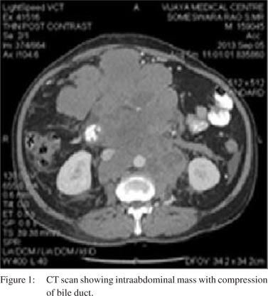
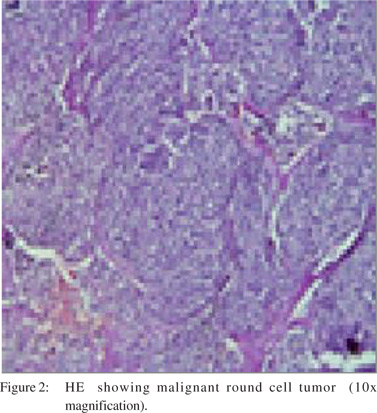
A 50-year-old male patient presented with back pain of 1 month’s duration. Abdominal examination revealed a palpable mass located in the lower abdomen. Routine laboratory tests were normal. USG abdomen was performed that showed multiple hypoechoic and heterogenous nodules in the peri-aortic and para-aortic regions. CT abdomen revealed a large lobulated solid mass (6.8 cm x 9.4 cm x 13.5 cm) in the right lumbar region extending into the right iliac fossa, multiple lymph nodes in the paraaortic, paracaval and retroperitoneal regions. Lymphoma was suspected and FNAC report was inconclusive. Biopsy showed round to polyhedral cells arranged in rosette pattern suggestive of cellular tumor mass and advised histological assessment. After 15 days he again presented to us with jaundice and worsening abdominal distention. Lab reports suggested obstructive jaundice. Repeat CT abdomen (Figure 1) showed large lobulated soft tissue mass (20 cm x 17 cm x16 cm) and there was enlargement of all groups of retroperitoneal and mesenteric lymph nodes, which was non-discrete. CBD was dilated (10 mm) and displaced anteriorly and compressed in its retro pancreatic segment by a large lymph nodal mass. Histology (Figure 2) was suggestive of malignant round cell tumor. Immunohistochemical studies included CD99 (+), synaptophysin (“), and LCA (-). In the context of clinical and histopathological evidence Ewing’s sarcoma was diagnosed in this patient.
Duodenoscopy showed duodenal encasement of tumor. Biopsy reported non-specific infiltration. Palliative chemotherapy with ifosfamide and etoposide was started.
Discussion
Extraosseous Ewing’s sarcoma (EES) occurs in the soft tissues (5%), often immediately adjacent to bones and is histologically indistinguishable from Ewing’s sarcoma.[2] Distinct membranous CD99 expression is characteristic and highly but non-specific reliable positive marker for EES.[3] ES, rhabdomyosarcoma, neuroblastoma, round cell liposarcoma are subcategories of small blue round cell tumors. Our first differential was lymphoma but biopsy revealed small round cell tumor, suggestive of EES. The mainstay of treatment should include multi-agent chemotherapy and aggressive surgical treatment.[5]
References
- Tefft M, Vawter GF, Mitus A. Paravertebral “round cell” tumors in children. Radiology. 1969;92:1501–9.
- Ahmad R, Mayol BR, Davis M, Rougraff BT. Extraskeletal Ewing’s sarcoma. Cancer. 1999;85:725–31.
- Mobley BC, Roulston D, Shah GV, Bijwaard KE, McKeever PE. Peripheral primitive neuroectodermal tumor/Ewing’s sarcoma of the craniospinal vault: case reports and review. Hum Pathol. 2006;37:845–53.
- Ludwig JA. Ewing sarcoma: historical perspectives, current stateof- the-art, and opportunities for targeted therapy in the future. Curr Opin Oncol. 2008;20:412–41.