|
|
|
|
 |
|
| |
 |
|
|
Case Report |
|
|
|
|
|
Keywords :
|
|
|
Sudhasmita Rauta, U Ramesh, Sesha Deepti, Pvb Ramlakshmi
Department of Pathology,
Maharaja Institute of Medical Science, Vizianagaram
NTR University, Andhra Pradesh, India
Corresponding Author:
Dr. Sudhasmita Rauta
Email: drsudhasmita@gmail.com
DOI:
http://dx.doi.org/10.7869/tg.190
48uep6bbphidvals|649 48uep6bbph|2000F98CTab_Articles|Fulltext Enterobius vermicularis is the most common helminth infecting man with a host range restricted to humans.[1] The parasite is not transmitted by arthropod vectors or intermediate hosts but directly from the parasitized subject to a healthy subject by means of infected hands, food or fomites. Indirect transmission by air has also been noted, wherein suspended microscopic eggs get inhaled with dust.[2] True granulomas caused by Enterobius vermicularis with their ova in internal genitalia, pelvis and appendix are well documented.[3-5] The lesion described here is unique in its location and clinical presentation.
Case report
A 33-year-old male presented to our surgical outpatient department with complaints of bleeding per rectum since on month. He also gave history of chronic intermittent diarrhoea. Per rectal digital examination revealed a firm tender mass in the anterior rectal wall. Proctoscopy showed second degree haemorrhoids and an irregular nodular mass 3 × 2 cm in size, in the anterior rectal wall.
Result of routine stool examination and barium study were normal. The mass was removed completely with a surgical diagnosis of either tuberculoma or carcinoma. Specimen sent for histopathology included two bits of grayish-white nodular tissue measuring 2.5 cm in diameter.
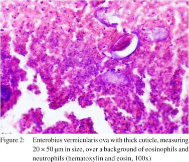
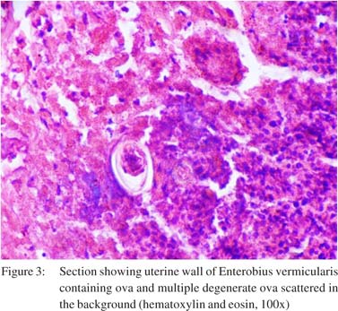
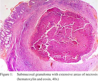 Histopathology revealed a submucosal granuloma extending up to the subserosa (Figure 1). Extensive areas of necrosis along with large number of eosinophils and neutrophils were observed in the periphery while an amorphous material occupied the centre containing numerous oval eggs bearing a thick cuticle and measuring approximately 20 × 50 µm in size. The ova were morphologically characteristic of E. vermicularis and were accompanied by the uterine wall remnants of the nematode (Figures 2 & 3).
 Discussion
We hypothesize that this unusual presentation in our case was caused by the aberrant migration of a gravid female E vermicularis nematode into the depths of the patient’s rectal wall. The worm eventually got trapped and died in the deadend fistula, releasing its ova, degradation products and inciting a local granulomatous inflammatory reaction.[6-9] E. vermicularis-induced granulomas can be asymptomatic or they may present with bleeding per rectum and peritoneal adhesions. The differential diagnosis of such lesions in these locations includes, sarcoidosis, tuberculosis, Crohn’s disease and foreign body reaction due talc, silicon or any surgical material.[10,11] Further, E. vermicularis cases of rectal granuloma have been rarely reported, as compared to appendicular and peri-anal lesion.[12]
Our report presents an unusual case of E. vermicularis granuloma and highlights the importance of considering helminthic parasitosis as a differential diagnosis for atypical gastrointestinal lesions.
References
Discussion
We hypothesize that this unusual presentation in our case was caused by the aberrant migration of a gravid female E vermicularis nematode into the depths of the patient’s rectal wall. The worm eventually got trapped and died in the deadend fistula, releasing its ova, degradation products and inciting a local granulomatous inflammatory reaction.[6-9] E. vermicularis-induced granulomas can be asymptomatic or they may present with bleeding per rectum and peritoneal adhesions. The differential diagnosis of such lesions in these locations includes, sarcoidosis, tuberculosis, Crohn’s disease and foreign body reaction due talc, silicon or any surgical material.[10,11] Further, E. vermicularis cases of rectal granuloma have been rarely reported, as compared to appendicular and peri-anal lesion.[12]
Our report presents an unusual case of E. vermicularis granuloma and highlights the importance of considering helminthic parasitosis as a differential diagnosis for atypical gastrointestinal lesions.
References
- Cram EB. Studies on oxyuriasis XXVIII. Summary and conclusions. Am J Dis Child. 1943;65:46–59.
- Hugot JP, Reinhard KJ, Gardner SL, Morand S. Human enterobiasis in evolution: origin, specificity and transmission. Parasite. 1999;6:201–8.
- Anthony PP, McAdam IW. Helminthic pseudotumours of the bowel: thirty-four cases of helminthoma. Gut. 1972;13:8–16.
- Aschoff L. Appendicopathia oxyurica. Med Klin. 1913;9:249-51. Quoted by Symmers W St C. Pathology of oxyuriasis. Arch Path. 1950;50:475–516.
- Chomet B. Oxyuris vermicularis infection of the wall offallopian tube. Arch Path.;34:742–4.
- Dundas KC, Calder AA, Alyusuf R. Enterobius vermicularis threadworm infestation of paraovarian tissue in a woman who has had a hysterectomy. Br J Obstet Gynaecol. 1999;106:605–7.
- Erhan Y, Zekioglu O, Ozdemir N, Sen S. Unilateral salpingitis due to enterobius vermicularis. Int J Gynecol Pathol. 2000;19:188–9.
- Knuth KR, Fraiz J, Fisch JA, Draper TW. Pinworm infestation of the genital tract. Am Fam Physician. 1988;38:127–30.
- Smolyakov R, Talalay B, Yanai-Inbar I, Pak I, Alkan M. Enterobius vermicularis infection of female genital tract: a report of three cases and review of literature. Eur J Obstet Gynecol Reprod Biol. 2003;107:220–2.
- McDonald GS, Hourihane DO. Ectopic Enterobius vermicularis. Gut. 1972;13:621–6.
- Bijlmer J. An exceptional case of oxyuriasis of the intestinal wall. J Parasitol. 1946;32:359–66.
- Avolio L, Avoltini V, Ceffa F, Bragheri R. Perianal granuloma caused by Enterobius vermicularis: report of a new observation and review of the literature. J Pediatr. 1998;132:1055–6.
|
|
|
|
|
|