48uep6bbphidvals|383
48uep6bbph|2000F98CTab_Articles|Fulltext
Introduction
Humanity’s battle against infectious diseases has continued since time immemorial. This tug of war between infective microorganisms and mankind has reached a stage where stakes are high and options for us are limited. Development of resistant micro-organisms and broad spectrum antibiotics used to treat them has further complicated the issue. It has led to resurgence of hospital acquired infections like Clostridium difficile which have considerable morbidity and mortality. Therefore, current strategies should focus on prevention rather than cure. Bacillus difficilis, an apparently innocuous organism, was isolated in 1935 from the fecal flora of healthy neonates.[1] Forty years later, it was renamed as Clostridium difficile (C. difficile) and identified as the cause of 15-20% of antibiotic–associated diarrhea (AAD) and all cases of pseudomembranous colitis (PMC).[2] It colonizes 3% of healthy adults[3], 20% hospitalized patients[4,5] and 25-80% of infants.[6] C.difficile is a Gram-positive, anaerobic spore-forming bacillus. C. difficile associated diarrhea (CDAD) was initially considered a “nuisance” disease as symptoms were mild, specific treatment was not always necessary (i.e. stopping antibiotic worked alone), treatmentwas cheap, sequelae were rare and usually not severe. Therefore, compared to other nosocomial diseases, this infection did not receive enough attention from researchers. This “nuisance” disease has lately become a “killer” disease with relatively higher mortality and morbidity.
What is the current knowledge?
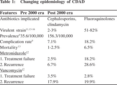
CDAD is now considered to be the commonest cause of nosocomial diarrhea. Its incidence and severity has dramatically increased since the appearance of a hypervirulent strain in 2000, especially in health care settings. It is estimated that CDAD results in more than 500,000 new cases of nosocomial diarrhea in United States annually, and results in 15,000 deaths per year.[7] It is estimated that each episode of C. difficile associated diarrhea increases 3.6 extra days of hospitalization and additional hospital costs of $3,669 per patient in the United States. Cumulative annual excess hospital healthcare cost due to C. difficile disease is estimated to be $1.1 billion in US.[8] This phenomenon is not confined only to United States; regional outbreaks of CDAD have been reported from world over.[9,10] An outbreak from Quebec in Canada was reported by Pepin et al[9] in which they reviewed 1721 patients with CDAD diagnosed between 1991 and 2003. The incidence of CDAD increased from 35.6 per 100,000 in 1991 to 156.3 per 100,000 in 2003. Moreover, among patients aged 65 years or more, the incidence increased from 102.0 to 866.5 per 100,000. The proportion of cases with CDAD that were complicated increased from 7.1% (12/169) in 1991–1992 to 18.2% (71/390) in 2003 (p < 0.001), and the mortality within 30 days after diagnosis increased from 4.7% (8/169) in 1991–1992 to 13.8% (54/390) in 2003 (p <0.001).[9] Similarly in England, C. difficile infection was the primary cause of death for 499 patients in 1999; its number rose to 3393 in 2006 and 3875 in 2007.[10]
Why there is an increase in the incidence of CDAD?
Emergence of hypervirulent strains coupled with indiscriminate use of antimicrobials and inadequate infection control measures in hospitals are primarily responsible factors for recent outbreaks of C. difficile infection. Molecular studies in C. difficile isolates from a number of US and Canadian healthcare centres have identified a more virulent strain of C. difficile. It is characterized as toxinotype III, North American Pulsed Field (NAP) type 1 and PCR-ribotype 027 (all synonymous terms) based on method of detection. This strain carries the binary toxin gene cdtB and an 18-bp deletion in tcdC gene from the pathogenicity locus. The tcdC gene is a putative regulatory gene that down-regulates transcription of tcdA and tcdB which encode toxin A and B, respectively. Deletion in tcdC gene results in loss of negative regulation in production of toxins A and B resulting in their increased production. The change from a relatively less virulent to more virulent isolate of C. difficile was substantiated by McDonald et al.[11] They compared 187 C. difficile isolates collected from outbreaks of CDAD between 2000 and 2003 with historical database of more than 6000 isolates obtained before 2001. BI/ NAP type 1 strain constituted more than 50 percent of isolates in current outbreaks as against only 14 cases in (more than 6000 patients) historical databases. All of the current, but none of the historic BI/NAP type 1 isolates were resistant to gatifloxacin and moxifloxacin (p<0.001).[11] Furthermore, Warny et al showed that peak median toxin A and toxin B concentrations produced in vitro by NAP type 1/027 were 16 and 23 times higher, respectively, than those measured in isolates representing 12 different PFGE types, known as toxinotype O.[12] Subsequently, it was established that that toxinotype III strain is associated with hospital outbreaks of increased disease severity of CDAD and a higher mortality.[13]
The distinct changes in the epidemiology of CDAD which have been observed are summarized in the Table 1.
Why it is important to us?
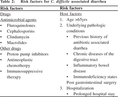
CDAD is under-recognized in India and Asia, due to lack of clinical suspicion, difficulty in culturing the organism and cost of toxin assay. Prevalence of CDAD is around 2 to 4% in patients without diarrhea and 7 to 30% in patients with diarrhea in different hospital based studies.[18,19,20,21,22,23] The prevalence has not been estimated in the community setting in India. The isolation of C. difficile was first described in 1985, when Gupta and Jadhav[18] isolated it from 25.3% of hospitalized patients with diarrhea and 4.3% of controls admitted for other ailments. Niyogi et al[19] isolated C. difficile in 11% hospitalized patients with diarrhea and 2.9% non-diarrheic controls; 87% isolates produced cytotoxin even though the diarrheic patients had no history of antibiotic use. Bhattacharya et al[20] investigated 233 patients with acute diarrhea and isolated C. difficile as a sole pathogen from 7.3%, of which, 82.4% produced cytotoxin. Vaishnavi et al[21] reported 30% positivity for C. difficile toxin in hospitalized patients of all age groups receiving single to multiple antibiotics for various diseases, but only in 7% of patients not receiving antibiotics. When only adult population were investigated, the positivity for C. difficile toxin was 19.4% in the antibiotic receiving hospitalized patients.[22] In our institution, C. difficile was found to be responsible for 15% of the cases of nosocomial diarrhea in 1999.[23] Majority of C. difficile isolates in India are responsive to metronidazole,[24,25] although there is a possibility that there may be emergence of resistant strains with severe disease manifestations in general practice. The risk factors associated with C.difficile associated diarrhoea are listed in Table 2.


How to define and categorize CDAD?
A CDAD case is defined as a case of diarrhea (i.e., un-formed stool that conforms to the shape of a specimen collection container) or toxic megacolon (i.e., abnormal dilation of the large intestine documented radiologically) without other known etiology that meets 1 or more of the following criteria: (1) a positive stool sample for C. difficile toxin A and/or B, demonstration of a toxin-producing C. difficile organism in the stool sample by culture or other means; (2) pseudomembranous colitis as seen during endoscopic examination or surgery; and (3) pseudomembranous colitis as seen during histopathological examination.[26]
A recurrent CDAD case is defined as an episode of CDAD (i.e., one that meets the criteria for a CDAD case) that occurs 8 weeks or less after the onset of a previous episode, provided that CDAD symptoms from the earlier episode resolved with or without therapy. The recurrent CDAD case definition may be implemented for laboratory-based reporting systems on the basis of the following stipulations: (1) an additional positive result of a laboratory test performed on a specimen collected 2 weeks or less after the last specimen that tested positive represents continuation of the same CDAD case, (2) an additional positive result of a laboratory test performed on a specimen collected 2-8 weeks after the last specimen that tested positive represents a recurrent CDAD case, and (3) an additional positive result of a laboratory test performed on a specimen collected more than 8 weeks after the last specimen that tested positive represents a new CDAD case.[26]
A case patient with severe CDAD is defined as a patient who meets any of the following criteria within 30 days after CDAD symptom onset (or, in the case of laboratory-based reporting, within 30 days after the index laboratory test): (1) history of admission to an intensive care unit for complications associated with CDAD (e.g., for shock that requires vasopressor therapy); (2) history of surgery (e.g., colectomy) for toxic megacolon, perforation, or refractory colitis; and (3) death caused by CDAD within 30 days after symptom onset (e.g., as listed on the death certificate or recorded in the medical record
by a clinician caring for the patient).[26]
A health care facility (HCF) is defined as any acute care, long-term care, long term acute care, or other facility in which skilled nursing care is provided and patients are admitted at least overnight. CDAD case patients are further defined by their exposures as follows:
A patient is classified as having HCF-onset, HCFassociated CDAD, when the onset of CDAD symptom is more than 48 hours after admission to an HCF.
A patient is classified as having community-onset, HCFassociated CDAD when the onset of CDAD symptom starts in the community or 48 hours or less after admission to an HCF, provided that symptom onset was less than 4 weeks after the last discharge from an HCF.
A patient is classified as having community-associated CDAD when the onset of the CDAD symptom starts in the community or 48 hours or less after admission to an HCF, provided that symptom onset was more than 12 weeks after the last discharge from an HCF. A patient is classified as having indeterminate CDAD when CDAD symptoms onset does not fit in any of the above criteria for an exposure setting. A patient is classified as having unknown CDAD when the exposure setting cannot be determined because of lack of available data.[26]
When to suspect?
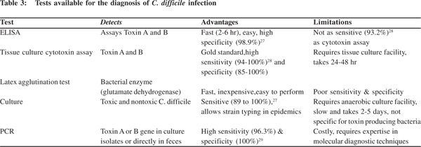
High index of suspicion is required for the diagnosis of C. difficile, especially when diarrhea occurs in hospital setting. Use of antibiotics complicated by diarrhea, fever, and leucocytosis are strong pointers towards a suspicion of CDAD. Clinical suspicion is usually confirmed by ELISA based toxin assays in stool sample. Other stool based tests have been elaborated in Table 3. Presentation with atypical manifestations may require colonoscopic examination and histological confirmation. There is no definite role of radiological investigations and they are not required unless there is a suspicion of complications such as toxic megacolon.
Management
The aim of therapy is to restore normal colonic microflora, resulting in the elimination of C. difficile. Patients with mild symptoms can occasionally be managed conservatively by discontinuation of the inciting antibiotic. It is effective in 15 to 25 % patients with no further need of any C. difficile specific antimicrobials.[30,31] However, in most patients anti-C. difficile therapy is required to ameliorate symptoms while allowing gradual restoration of the normal gut flora. Confirmation of diagnosis with fecal toxin assay is essential prior to initiating antibiotic therapy as symptoms of this condition are nonspecific. Initiation of empirical treatment can be considered in acutely ill patients awaiting test result or if there is strong suspicion of C. difficile infection. In most patients, sigmoidoscopic or colonoscopic examination is not required for establishing the diagnosis, as pseudomembranes occur in only half of infected patients, moreover these procedures carry a higher risk of complication including perforation.[32] The specific indications for colonoscopic examinations include a need for early diagnosis of CDAD and stool test is delayed or unavailable; and when other colonic diseases are in differential diagnosis.

Antibiotic treatment for initial C. difficile infection
Treatment of C. difficile infection needs to be individualized depending on the severity of the disease and patient characteristics. The predictors of severe disease include severe diarrhea, ileus, fever, age more than 60 years, acute renal failure, hypoalbuminemia, ICU stay and pseudomembranes seen on colonoscopy.
Majority of patients will require antibiotic therapy and, whenever possible, discontinuation of the predisposing antibiotics. Patients who continue on implicated antibiotics while undergoing treatment of CDAD have a higher likelihood of treatment failure.33 Younger patients with mild symptoms might respond to discontinuation of offending antibiotic and hydration alone. In fact, non use of antibiotic is associated with faster elimination of spores in the carriers of C. difficile.
Presence of co-morbidities and features of severe infection calls for antibiotic therapy. Despite initial response rates of >90%;[34,35] 15%–30% of patients have relapse of infection after successful initial therapy, usually in the first few weeks after treatment is discontinued.[2,36] Recurrence may involve re- infection with the initial strain or a new strain. Failure to develop specific antibody response has recently been identified as a critical factor for recurrence of infection.[37]
Metronidazole and vancomycin are the mainstay of the treatment of CDAD, as both these agents are highly active against all strains of pathogenic C. difficile. Neither of these drugs is however effective for C. difficile carriage. They also have no role in prevention of C. difficile infection while patients are receiving other antimicrobials.[38] Other antibiotics (bacitracin, teicoplanin, fusidic acid,[39] nitazoxanide[40], rifaximin[41], ramoplanin[42]) have been tried as a single or adjuvant agent in CDAD with variable efficacy. Metronidazole is currently used as first-line therapy because it is cheap, easily available and has reasonable efficacy. The standard initial therapy is 500 mg orally (7.5–15 mg/kg in children) 3 times daily for 14 days, which successfully resolves symptoms in >90% of patients within 1 week.[34,35] Parenteral or rectaladministration can be used in patients who are unable to take orally and leads to similar systemic and colonic tissue drug levels. Metronidazole preferentially diffuses from the serum and interstitial compartment of the inflamed colon into the colonic lumen, a property that can increase its efficacy againstC. difficile.[43] The side effects of metronidazole though relatively frequent and unpleasant but are rarely serious and do not require discontinuation in most instances. Anorexia, nausea, metallic taste and abdominal cramps are the most common side effects. Rarely, neutropenia, peripheral neuropathy and seizure may result. The use of metronidazole should be avoided in neurological diseases, blood dyscrasias,first trimester of pregnancy and in those who are chronic alcoholics.
Based on a recent guideline[44] and Cochrane review,[45] oral vancomycin is recommended as first-line therapy in pregnant women, in children younger than 10 years of age, and those with severe infections. Oral vancomycin should also be used for patients where either metronidazole is contraindicated or has failed to show response. It is a tricyclic glycopeptides bactericidal antibiotic active against Gram positive bacteria. It is administered orally in C. difficile infection. It is not absorbed in the small intestine because of its large molecular weight and reaches the colon intact, an advantage for the treatment of C.
difficile. Parenteral vancomycin is not effective as it is not excreted into the colonic lumen. Unfortunately, oral vancomycin is not available in India. The recommended dose to treat C.
difficile for the first episode of infection and first recurrence is 125 mg 4 times daily for 10–14 days. As it is not absorbed systemically, side effects such as nephrotoxicity, ototoxicity and red-man syndrome are not seen which otherwise are well known with parenteral vancomycin. Nausea and mild abdominal discomfort are the most common side effects of oral vancomycin and rarely limit treatment. Vancomycin enemas can be used when oral administration is not feasible. Colonoscopic decompression with vancomycin lavage is reported to be useful in one study.[46]
Treatment of acute fulminant infection
Severe C. difficile colitis tends to occur during the initial infection or first recurrence. It is associated with high morbidity and estimated case–fatality rate is about 2%.[47] The risk factors for development of acute fulminant infection include advanced age, presence of co-morbidities, high total leucocyte count, and lactic acidosis. Patients with toxic megacolon may not have diarrhea. Hemodynamic instability is consistently associated with high rates of mortality.
Medical management of fulminant disease should include administration of both vancomycin orally and metronidazole intravenously. Because of ileus, if oral administration of vancomycin is not possible vancomycin should be administered rectally. Patients not responding to above regimen may require urgent surgical intervention. In patients who are at surgical risk can be given pooled human immunoglobulin (200–500 mg/kg/day) until they improve, although evidence to support its efficacy is lacking.[48]
Immediate surgical intervention is indicated for patients with ileus, toxic megacolon, or those having localizing peritoneal signs. Subtotal colectomy is the procedure of choice. Though prompt subtotal colectomy and ileostomy is generally advised, it carries a high (35% to 80%) mortality rate. Hemicolectomy should be avoided as it is associated with higher mortality.[49]
Treatment strategy for recurrent C. difficile infection Approximately 15%-30% of patients experience a symptomatic recurrence after discontinuation of antibiotics.[2,36]
Approximately half of the recurrent C. difficile infections are caused by repeated ingestion of bacteria (re-infection) before the normal intestinal flora recovers, and half are attributed to incomplete eradication of the original strain during antibiotic treatment (relapse).[50] Older patients and those with a history of prior C. difficile recurrence are at an even higher risk (>50%)
Clostridium difficile associated diarrhea 19
for recurrent disease. Most recurrences happen within 2 weeks of stopping therapy, but delayed recurrences have also been documented. The first recurrence can be treated with the same antibiotic, for example a 14-day course of metronidazole (if tolerated) or with supportive care alone, if symptoms are mild. Unfortunately, those who have recurrence of the disease have higher chances (up to 65%) of further recurrences.[51]
Management strategies for recurrent C. difficile infection include repeat courses of metronidazole or vancomycin, tapered and pulsed dosing of vancomycin, toxin-binding agents such as cholestyramine, probiotics such as Saccharomyces boulardii, and immunotherapy, but none are supported by randomized
large clinical trials.
For patients who experience more than one recurrences of symptomatic C. difficile infection, the diagnosis of recurrent C. difficile infection should be confirmed by a stool assay before starting therapy. For confirmed recurrent C. difficile infection, a 6-week pulsed-tapered (125 mg qid × 10-14 d; 125 mg bid × 7 d; 125 mg daily × 7 d; 125 mg once every 2 d × 8 d; and 125 mg once every 3 d × 15 d) course of oral vancomycin therapy can be tried.[52] Pulsed dosing of antibiotics allows remnant C. difficile spores to germinate into antibiotic-sensitive vegetative forms, which are then killed on subsequent day of antibiotic therapy. It also restores colonic microflora while conferring adequate protection against C. difficile.[53] Toxinbinding resin (colestipol 5 g every 12 hours) may provide additional benefit when added in the later weeks of the antibiotic taper and continued for 4–6 weeks after antibiotic therapy is stopped.[54] Addition of rifaximin (400–800 mg daily) in 2 or 3 divided doses for 2 weeks, immediately after 10–14 days of vancomycin therapy while the patient is still in clinical remission has been found to be an effective strategy for those who have multiple relapses of CDAD.[41]
Despite these treatment strategies, if the patient continues to have recurrent CDAD, chronic vancomycin therapy at the lowest effective dose (125 mg daily or every alternate day) can be tried for an indefinite period. Probiotics, immunotherapy, and fecal bacteriotherapy are other options if recurrences are frequent and severe.
Probiotics are beneficial bacteria that modulate mucosal and systemic immunity and contribute to microbial equilibrium in human gut. Therefore, they have been advocated for antibiotic associated and CDAD which results from alteration of normal fecal flora. However, evidence to support routine use of probiotics for CDAD is insufficient and conflicting. A recent Cochrane review failed to demonstrate its efficacy in treatment of CDAD.[55] Some of the probiotic strains(Saccharomyces boulardii) can be used in recurrent CDAD either with metronidazole or high dose vancomycin.[56,57] The role of non-toxigenic strain of C. difficile as probiotic therapy was explored in two patients with relapsing C. difficile diarrhea and found to be effective.[58] There are evidences to support role of few probiotic strains (Saccharomyces boulardii, Lactobacillus rhamnosus GG) in the prevention of antibiotic associated diarrhea, but data on their role in prevention of CDAD is sparse.[59]
Fecal bacteriotherapy restores colon homeostasis by reintroducing missing bacterial flora in stool obtained from a healthy donor usually in form of enemas. It is effective (94- 100% success), safe, inexpensive and simple to administer in most hospitals.[60] However potential risk of transmitting infectious organism coupled with medico-legal problems and its esthetically unappealing nature has limited its role in clinical practice.
Asymptomatic carriers of C. difficile have been found to have 3 fold higher IgG antitoxin-A antibody compared with symptomatic patients.[61] Patients with higher IgG antitoxin-A antibody response during first episode of C. difficile infection have markedly (48 times) less risk of developing recurrent C. difficile infection than those with low antibody titers.[37] These two evidences are the basic principles for genesis of a new form of therapy for recurrent CDAD. Intravenous infusion of normal pooled human immunoglobulin (IVIG 400 mg/kg body weight) increases serum IgG antitoxin concentrations, therefore IV immunoglobulin has been used with variable success to treat a small number of patients with severe C. difficile colitis. However, IVIG failed to demonstrate any significant differences in mortality, colectomies and length of hospital stay in a retrospective review of 18 patients where it was used.[62]
What to do for asymptomatic carriers?
Universal precautions should be instituted for all patients in hospital to reduce the nosocomial spread of infection, as asymptomatic carriers act as hidden reservoir for C. difficile. Treatment of asymptomatic carriers is not warranted as it is likely to prolong the carrier state.[63] They are also resistant to acquisition of outbreak-associated strains[64] and no more likely to develop CDAD than those with negative stool cultures.[65,66] Further, data is presently inadequate to show reduction in hospital spread of infection following treatment of asymptomatic carriers.[38]
How to prevent C. difficile infection?
Prevention of C. difficile infection has two major components:
prevention of spread of organism to the patients and b) reduction in likelihood of clinical disease.
a) Prevention of spread of organism to the patients
It involves proper hand hygiene, use of personal protective equipment, environmental decontamination, isolation or cohort nursing and adequate treatment of CDAD cases. Hand washing is of utmost importance in prevention of hospital acquired infections. Vigilant hand hygiene consists of hand washing with friction for at least 15 seconds.[67] Alcohol based hand gels are highly effective against non-spore forming organisms, but they are not sporicidal. Due to the theoretical advantage of hand washing over alcohol-based hand sanitizers, hand washing with a non-antimicrobial soap or antimicrobial soap and water should be considered after removing gloves in the setting of a CDAD outbreak or if ongoing transmission cannot be controlled by other measures. UK national guidelines recommend healthcare workers to wash their hands before and after contact with patients with suspected or confirmed C. difficile infection. Protective disposable gloves and aprons should be used while handling body fluids and nursing CDAD patients.[68]
C. difficile spores can survive in the environment for months or years, and environmental contamination has been linked to the spread of C. difficile infection in healthcare settings. UK national guidelines therefore recommend cleaning of rooms, bed spaces, commodes, toilets and bathrooms of infected patients with chlorine containing cleaning agents or vaporised hydrogen peroxide daily (as they inactivate C. difficile spores). Use of disposable thermometers instead of electronic oral and rectal thermometers is recommended.[68]
Isolation or cohorting of patients with CDAD and enteric or contact precautions have been successful in limiting transmission of C. difficile in hospitals.[69,70] However, it is not possible to determine the specific effectiveness of isolation techniques as these measures are instituted as part of other infection control interventions.
Surveillance of C. difficile infections should be instituted to identify outbreaks and initiate control measures in a timely fashion. Early identification and treatment of CDAD cases prevent further spread of infection. No treatment is required for the asymptomatic carriers of C. difficile.
b) Reduction in likelihood of clinical disease
Antimicrobials should be prescribed according to local policies and guidelines for treatment and prophylaxis. Restrictions in the use of broad-spectrum antibiotics have shown to reduce the frequency of CDAD. Prospective observational cohort studies suggest that restricted use of clindamycin and third generation cephalosporins result in fewer cases of C. difficile infection.[71,72]
Besides antibiotics, use of has proton pump inhibitors has been associated with C. difficile infection, according to a recent meta-analysis involving 33,193 patients. Therefore, indiscriminate and unwarranted use of proton pump inhibitor(s) should be avoided specially in health care setting.[73]
Conclusions
The incidence of health care associated CDAD is on the rise with associated increased morbidity and mortality. C. difficile associated diseases are not commonly recognized in many health care settings. Atypical hypervirulent BI/NAP1/027 strain is resistant to flouroquinolones, produces more toxins and carries the gene encoding binary toxin which has led to increased incidence and severity of CDAD. Vancomycin and metronidazole remain the mainstay of treatment for CDAD, but newer investigational therapies hold promise. Control of health care associated CDAD involves a range of primary preventive measures.
Learning points
· Suspect CDAD when diarrhea occurs in appropriate clinical setting (use of antibiotic in the last 2 months or onset of diarrhea within 48 hrs or more after hospitalization).
· Pay careful attention to the supportive care (such as fluid and electrolyte replacement).
· Stop prescribing antibiotics if possible, and if not, substitute with a lower risk agent.
· Use metronidazole for initial treatment of patients with mild or moderate disease, and reserve vancomycin for severe disease.
· Isolate patients with suspected C. difficile infection, use gowns and gloves when seeing them, and remember to wash hands rather than use alcohol hand rubs.
· Retreat first-time recurrences with the same regimen used to treat the initial episode.
Clostridium difficile associated diarrhea 21
References
1. Hall IC, O’Toole E. Intestinal flora in new-born infants with a description of a new pathogenic anaerobe, Bacillus difficilis. Am J Dis Child. 1935;49:390–402.
2. Kelly CP, Pothoulakis C, LaMont JT. Clostridium difficile colitis. N Engl J Med. 1994;330:257–62.
3. Djuretic T, Wall PG, Brazier JS. Clostridium difficile: an update on its epidemiology and role in hospital outbreaks in England and Wales. J Hosp Infect. 1999;41:213–8.
4. McFarland LV, Mulligan ME, Kwok RY, Stamm WE. Nosocomial acquisition of Clostridium difficile infection. N Engl J Med. 1989;320:204–10.
5. Samore MH, DeGirolami PC, Tlucko A, Lichtenberg DA, Melvin ZA, Karchmer AW. Clostridium difficile colonization and diarrhea at a tertiary care hospital. Clin Infect Dis. 1994;18:181–7.
6. Kelly CP, LaMont JT. Clostridium difficile infection. Annu Rev Med. 1998;49:375–90.
7. McDonald LC, Owings M, Jernigan DB. Clostridium difficile infection in patients discharged from US short-stay hospitals, 1996-2003. Emerg Infect Dis. 2006;12:409–15.
8. Kyne L, Hamel MB, Polavaram R, Kelly CP. Health care costs and mortality associated with nosocomial diarrhea due to Clostridium difficile. Clin Infect Dis. 2002;34:346–53.
9. Pépin J, Valiquette L, Alary ME, Villemure P, Pelletier A, Forget K, et al. Clostridium difficile-associated diarrhea in a region of Quebec from 1991 to 2003: a changing pattern of disease severity. CMAJ. 2004;171:466–72.
10. Office for national statistics, UK statistics authority [homepage on the internet]. Newport, United Kingdom: [updated 2011 Mar 11; cited 2010 Sep 15]. Deaths involving Clostridium difficile, England and Wales, 1999–2009. Available from: http:// www.statistics.gov.uk/statbase/Product.asp?vlnk=14782
11. McDonald LC, Killgore GE, Thompson A, Owens RC Jr, Kazakova SV, Sambol SP, et al. An epidemic, toxin gene-variant strain of Clostridium difficile. N Engl J Med. 2005;353:2433–41.
12. Warny M, Pepin J, Fang A, Killgore G, Thompson A, Brazier J, et al. Toxin production by an emerging strain Clostridium difficile associated with outbreaks of severe disease in North America and Europe. Lancet. 2005;366:1079–84.
13. Loo VG, Poirier L, Miller MA, Oughton M, Libman MD, Michaud S, et al. A predominantly clonal multi-institutional outbreak of Clostridium difficile-associated diarrhea with high morbidity and mortality. N Engl J Med. 2005;353:2442–9.
14. Rupnik M, Avesani V, Janc M, von Eichel-Streiber C, Delmée M. A novel toxinotyping scheme and correlation of toxinotypes with serogroups of Clostridium difficile isolates. J Clin Microbiol.1998;36:2240–7.
15. Rupnik M, Brazier JS, Duerden BI, Grabnar M, Stubbs SL. Comparison of toxinotyping and PCR ribotyping of Clostridium difficile strains and description of novel toxinotypes. Microbiology. 2001;147:439–47.
16. Geric B, Rupnik M, Gerding DN, Grabnar M, Johnson S. Distribution of Clostridium difficile variant toxinotypes and strains with binary toxin genes among clinical isolates in an American hospital. J Med Microbiol. 2004;53:887–94.
17. Kelly CP, LaMont JT. Clostridium difficile—more difficult than ever. N Engl J Med. 2008;359:1932–40.
18. Gupta U, Yadav RN. Clostridium difficile in hospital patients. Indian J Med Res. 1985;82:398–401.
19. Niyogi SK, Bhattacharya SK, Dutta P, Naik TN, De SP, Sen D, et al. Prevalence of Clostridium difficile in hospitalised patients with acute diarrhoea in Calcutta. J Diarrhoeal Dis Res. 1991;9:16–9.
20. Bhattacharya MK, Niyogi SK, Rasaily R, Bhattacharya SK, Dutta P, Nag A, et al. Clinical manifestation of Clostridium difficile enteritis in Calcutta. J Assoc Physicians India. 1991;39:683–4.
21. Vaishnavi C, Kochhar R, Bhasin DK, Thapa BR, Singh K. Detection of Clostridium difficile toxin by an indigenously developed latex agglutination assay. Trop Gastroenterol.1999;20:33–5.
22. Vaishnavi C, Bhasin D, Kochhar R, Singh K. Clostridium difficile toxin and faecal lactoferrin assays in adult patients. Microbes Infect 2000;2:1827–30.
23. Dhawan B, Chaudhry R, Sharma N. Incidence of Clostridium difficile infection: a prospective study in an Indian hospital. J Hosp Infect. 1999;43:275–80.
24. Chaudhry R, Joshy L, Kumar L, Dhawan B. Changing pattern of Clostridium difficile associated diarrhoea in a tertiary care hospital: a 5 year retrospective study. Indian J Med Res. 2008;127:377–82.
25. Niyogi SK. Antimicrobial susceptibility of Clostridium difficile strains isolated from hospitalised patients with acute diarrhoea. J Diarrhoeal Dis Res. 1992;10:156–8.
26. McDonald LC, Coignard B, Dubberke E, Song X, Horan T, Kutty PK; Ad Hoc Clostridium difficile Surveillance Working Group. Recommendations for surveillance of Clostridium difficileassociated disease. Infect Control Hosp Epidemiol. 2007;28:140–5.
27. Lyerly DM, Neville LM, Evans DT, Fill J, Allen S, Greene W, et al. Multicenter evaluation of the Clostridium difficile TOX A/B TEST. J Clin Microbiol. 1998;36:184–90.
28. Sunenshine RH, McDonald LC. Clostridium difficile-associated disease: new challenges from an established pathogen. Cleve Clin J Med. 2006;73:187–97.
29. Alonso R, Muñoz C, Gros S, García de Viedma D, Peláez T, Bouza E. Rapid detection of toxigenic Clostridium difficile from stool samples by a nested PCR of toxin B gene. J Hosp Infect. 1999;41:145–9.
30. Olson MM, Shanholtzer CJ, Lee JT Jr, Gerding DN. Ten years of prospective Clostridium difficile-associated disease surveillance and treatment at the Minneapolis VA Medical Center, 1982– 1991. Infect Control Hosp Epidemiol. 1994;15:371–81.
31. Teasley DG, Gerding DN, Olson MM, Peterson LR, Gebhard RL, Schwartz MJ, et al. Prospective randomised trial of metronidazole versus vancomycin for Clostridium-difficileassociated diarrhoea and colitis. Lancet. 1983;2:1043–6.
32. Bartlett JG, Gerding DN. Clinical recognition and diagnosis of Clostridium difficile infection. Clin Infect Dis. 2008;46:S12–8.
33. Modena S, Gollamudi S, Friedenberg F. Continuation of antibiotics is associated with failure of metronidazole for Clostridium difficile-associated diarrhea. J Clin Gastroenterol. 2006;40:49–54.
34. Fekety R, Silva J, Kauffman C, Buggy B, Deery HG. Treatment of antibiotic-associated Clostridium difficile colitis with oral vancomycin: comparison of two dosage regimens. Am J Med. 1989;86:15–9.
35. Wilcox MH, Howe R. Diarrhoea caused by Clostridium difficile: response time for treatment with metronidazole and vancomycin. J Antimicrob Chemother. 1995;36:673–9.
36. Fekety R. Guidelines for the diagnosis and management of Clostridium difficile-associated diarrhea and colitis. American College of Gastroenterology, Practice Parameters Committee. Am J Gastroenterol. 1997;92:739–50.
37. Kyne L, Warny M, Qamar A, Kelly CP. Association between antibody response to toxin A and protection against recurrent Clostridium difficile diarrhoea. Lancet. 2001;357:189–93.
38. Gerding DN, Muto CA, Owens RC Jr. Treatment of Clostridium difficile infection. Clin Infect Dis. 2008;46:S32–42.
39. Zimmerman MJ, Bak A, Sutherland LR. Review article: treatment of Clostridium difficile infection. Aliment Pharmacol Ther. 1997;11:1003–12.
40. Musher DM, Logan N, Mehendiratta V, Melgarejo NA, Garud S, Hamill RJ. Clostridium difficile colitis that fails conventional metronidazole therapy: response to nitazoxanide. J Antimicrob Chemother. 2007;59:705–10.
41. Johnson S, Schriever C, Galang M, Kelly CP, Gerding DN. Interruption of recurrent Clostridium difficile-associated diarrhea episodes by serial therapy with vancomycin and rifaximin. Clin Infect Dis. 2007;44:846–8.
42. Peláez T, Alcalá L, Alonso R, Martín-López A, García-Arias V, Marín M, et al. In vitro activity of ramoplanin against Clostridium difficile, including strains with reduced susceptibility to vancomycin or with resistance to metronidazole. Antimicrob Agents Chemother. 2005;49:1157–9.
43. Bolton RP, Culshaw MA. Faecal metronidazole concentrations during oral and intravenous therapy for antibiotic associated colitis due to Clostridium difficile. Gut. 1986;27:1169–72.
44. Gerding DN, Johnson S, Peterson LR, Mulligan ME, Silva J Jr. Clostridium difficile-associated diarrhea and colitis. Infect Control Hosp Epidemiol. 1995;16:459–77.
45. Nelson R. Antibiotic treatment for Clostridium difficile-associated diarrhea in adults. Cochrane Database Syst Rev. 2007;3:CD004610.
46. Shetler K, Nieuwenhuis R, Wren SM, Triadafilopoulos G. Decompressive colonoscopy with intracolonic vancomycin administration for the treatment of severe pseudomembranous colitis. Surg Endosc. 2001;15:653–9.
47. Zilberberg MD, Shorr AF, Kollef MH. Increase in adult Clostridium difficile-related hospitalizations and case-fatality rate, United States, 2000–2005. Emerg Infect Dis. 2008;14:929–31.
48. Leffler DA, Lamont JT. Treatment of Clostridium difficileassociated disease. Gastroenterology. 2009;136:1899–912.
49. Koss K, Clark MA, Sanders DS, Morton D, Keighley MR, Goh J. The outcome of surgery in fulminant Clostridium difficile colitis. Colorectal Dis. 2006;8:149–54.
50. Wilcox MH, Fawley WN, Settle CD, Davidson A. Recurrence of symptoms in Clostridium difficile infection—relapse or reinfection? J Hosp Infect. 1998;38:93–100.
51. McFarland LV, Elmer GW, Surawicz CM. Breaking the cycle: treatment strategies for 163 cases of recurrent Clostridium difficile disease. Am J Gastroenterol. 2002;97:1769–75.
52. Kyne L, Kelly CP. Recurrent Clostridium difficile diarrhoea. Gut. 2001;49:152–3.
53. Tedesco FJ, Gordon D, Fortson WC. Approach to patients with multiple relapses of antibiotic-associated pseudomembranous colitis. Am J Gastroenterol. 1985;80:867–8.
54. Tedesco FJ. Treatment of recurrent antibiotic-associated pseudomembranous colitis. Am J Gastroenterol. 1982;77:220–1.
55. Pillai A, Nelson R. Probiotics for treatment of Clostridium difficile-associated colitis in adults. Cochrane Database Syst Rev. 2008;1:CD004611.
56. McFarland LV, Surawicz CM, Greenberg RN, Fekety R, Elmer GW, Moyer KA, et al. A randomized placebo-controlled trial of Saccharomyces boulardii in combination with standard antibiotics for Clostridium difficile disease. JAMA. 1994;271:1913–8.
57. Surawicz CM, McFarland LV, Greenberg RN, Rubin M, Fekety R, Mulligan ME, et al. The search for a better treatment for recurrent Clostridium difficile disease: use of high-dose vancomycin combined with Saccharomyces boulardii. Clin Infect Dis. 2000;31:1012–7.
58. Seal D, Borriello SP, Barclay F, Welch A, Piper M, Bonnycastle M. Treatment of relapsing Clostridium difficile diarrhoea by administration of a non-toxigenic strain. Eur J Clin Microbiol. 1987;6:51–3.
59. McFarland LV. Meta-analysis of probiotics for the prevention of antibiotic associated diarrhea and the treatment of Clostridium difficile disease. Am J Gastroenterol. 2006;101:812–22.
60. Aas J, Gessert CE, Bakken JS. Recurrent Clostridium difficile colitis: case series involving 18 patients treated with donor stool administered via a nasogastric tube. Clin Infect Dis. 2003;36:580–5.
61. Kyne L, Warny M, Qamar A, Kelly CP. Asymptomatic carriage of Clostridium difficile and serum levels of IgG antibody against toxin A. N Engl J Med. 2000;342:390–7.
62. Juang P, Skledar SJ, Zgheib NK, Paterson DL, Vergis EN, Shannon WD, et al. Clinical outcomes of intravenous immune globulin in severe clostridium difficile-associated diarrhea. Am J Infect Control. 2007;35:131–7.
63. Johnson S, Homann SR, Bettin KM, Quick JN, Clabots CR,Peterson LR, et al. Treatment of asymptomatic Clostridium difficile carriers (fecal excretors) with vancomycin or metronidazole. A randomized, placebo-controlled trial. Ann Intern Med. 1992;117:297–302.
64. Samore MH. Epidemiology of nosocomial clostridium difficile diarrhoea. J Hosp Infect. 1999;43:S183–90.
65. Johnson S, Clabots CR, Linn FV, Olson MM, Peterson LR, Gerding DN. Nosocomial Clostridium difficile colonisation and disease. Lancet. 1990;336:97–100.
66. Shim JK, Johnson S, Samore MH, Bliss DZ, Gerding DN. Clostridium difficile associated diarrhea 23 Primary symptomless colonisation by Clostridium difficile and decreased risk of subsequent diarrhoea. Lancet. 1998;351:633–6.
67. Boyce JM, Pittet D; Healthcare Infection Control Practices Advisory Committee. Society for Healthcare Epidemiology of America. Association for Professionals in Infection Control. Infectious Diseases Society of America. Hand Hygiene Task Force. Guideline for Hand Hygiene in Health-Care Settings: recommendations of the Healthcare Infection Control Practices Advisory Committee and the HICPAC/SHEA/APIC/IDSA Hand Hygiene Task Force. Infect Control Hosp Epidemiol. 2002;23:S3–40.
68. Department of Health and Health Protection Agency [homepage on the internet]. United Kingdom: Health Protection Agency; c2011 [updated 2010 Jan 26; cited 2010 Sep 15]. Clostridium difficile infection: how to deal with the problem. Available from: www.hpa.org.uk/webw/HPAweb&HPAwebStandard/HPAweb_C/ 1204186173530
69. Struelens MJ, Maas A, Nonhoff C, Deplano A, Rost F, Serruys E, et al. Control of nosocomial transmission of Clostridium difficile based on sporadic case surveillance. Am J Med. 1991;91:138S–144S.
70. Zafar AB, Gaydos LA, Furlong WB, Nguyen MH, Mennonna PA. Effectiveness of infection control program in controlling nosocomial Clostridium difficile. Am J Infect Control. 1998;26:588–93.
71. Khan R, Cheesbrough J. Impact of changes in antibiotic policy on Clostridium difficile-associated diarrhoea (CDAD) over a fiveyear period in a district general hospital. J Hosp Infect. 2003;54:104–8.
72. Davey P, Brown E, Fenelon L, Finch R, Gould I, Hartman G, et al. Interventions to improve antibiotic prescribing practices for hospital inpatients. Cochrane Database Syst Rev.2005;4:CD003543.
73. Shukla S, Shukla A, Guha S, Mehboob S. Use of proton pump inhibitors and risk of Clostridium difficile-associated diarrhea: a meta-analysis. Gastroenterology. 2010;138(Suppl 1):S209.