|
|
|
|
 |
|
| |
 |
|
|
Case Report |
|
|
|
|
|
Keywords :
|
|
|
N Jain, Cv Kantharia, Ry Prabhu, Padma*, An Supe, Rd Bapat
Department of Surgical Gastroenterology and Radiology*
Seth GSMC & KEM Hospital
Mumbai, 400012, India.
Corresponding Author:
Dr Chetan V Kantharia
Email: kanthariachetan@yahoo.com
DOI:
http://dx.doi.org/
48uep6bbphidvals|284 48uep6bbphidcol4|ID 48uep6bbph|2000F98CTab_Articles|Fulltext Duodenal diverticula are relatively infrequent. They are usually managed conservatively, until complications such as diverticulitis, upper gastrointestinal bleeding, perforation, biliopancreatic obstruction and duodenal obstruction occur.[1] Abdominal pain is the most common presentation of duodenal diverticulum.[2] We report two cases of diverticula, one each of the true and pseudo varieties, arising from the third part of the duodenum. While one patient presented with acute pain, the other did so with chronic pain in the abdomen. Both patients were treated surgically.
Case 1
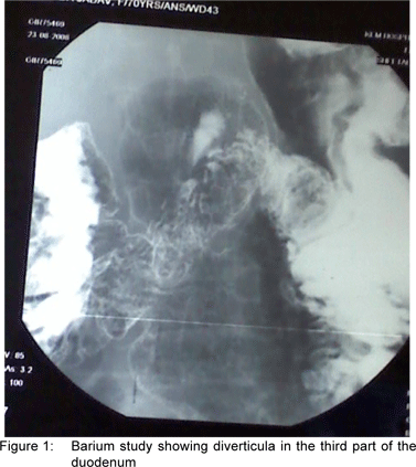
A 76-year-old lady, known case of bronchial asthma and hypertension was referred to us from another hospital with acute abdomen and a differential diagnosis of acute cholecystitis/ acute pancreatitis. Her clinical history revealed epigastric pain, and episodic nausea and vomiting over the last four years. She had been hospitalised previously on two occasions and treated symptomatically. The abdominal examination was unremarkable. Investigations revealed a normal HIDA scan, normal CT scan with sludge in the gallbladder and an upper gastrointestinal (GI) endoscopy which was normal till the second part of the duodenum. BMFT study revealed diverticula in the third part of the duodenum.(Figure 1) She was managed conservatively.
Four weeks later, she was taken up for surgery and a diverticulum arising from the anti-mesenteric border of the third part of the duodenum with adhesions was seen. (Figure 2) The duodenum was mobilised with Kocher‘s manoeuvre. By a combination of blunt and sharp dissection the diverticulum was isolated and defined. Diverticulectomy was performed using a 55 mm linear cutter to resect the base of the diverticulum. Posterior gastrojejunostomy was done to protect the duodenal suture line and divert food. The postoperative period was uneventful. Histopathology reported a 5.6 X 4.3 cm size diverticulum with normal duodenal mucosa, submucosal and congested serosa suggesting a true diverticulum.
Case 2
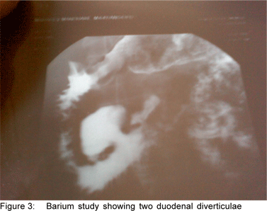
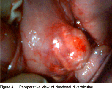
A 67-year-old male patient was admitted with a two-year history of non-colicky, dull aching abdominal pain in the upper abdomen associated with postprandial fullness. He was being treated symptomatically for these complaints. On examination he was pale and the abdominal examination was unremarkable. Upper GI endoscopy, colonoscopy and hematology were normal. The CT scan showed a diverticulum in the third part of the duodenum. BMFT study revealed two diverticula in the third part, the larger of the two was 4.4 cm and the smaller was 1.5 cm in length.(Figure 3) Having ruled out all other causes of upper abdominal pain, the patient was taken up for surgery. The duodenum was mobilised with Kocher‘s manoeuvre. The diverticulum was identified, dissected isolated and defined.(Figure 4) Due to dense adhesions, this was difficult as compared to the first case. Mucosal out-pouching was noticed in the meso-duodenal region of the third part of the duodenum. As it was a pseudodiverticulum, it was inverted. The postoperative period was uneventful and patient was symptom-free.
Discussion
The duodenum is the commonest site of small bowel diverticula.[3] Though most of the diverticula occur in the second part of the duodenum, especially near the ampulla of Vater,[4] the diverticulum of the third part of the duodenum as seen in our patients is not so common. The incidence of duodenal diverticula varies from 2.5-3% in upper GI series and 15-22% in autopsies.[5] 85-90% are asymptomatic and 10-15% symptomatic with only 1-2% requiring surgery.[6] The size of diverticula usually vary from 1-3 cm. Giant diverticula occur on the anti-mesenteric side as growth of the diverticulum is restricted by the pancreas on the medial side. Diverticula are of two types- Pseudodiverticulum which is a mucosal outpouching arising from the duodenal wall where the vessel penetrates; and true diverticulum which comprises all layers of the duodenum and considered congenital in origin.[7] Pseudodiverticula are more common than true diverticula. Duodenal diverticula can be extra-luminal or intra-luminal; the former is more common. Intra-luminal diverticula are usually associated with congenital anomalies like web and atresia of duodenum. Duodenal diverticula are asymptomatic until complications such as diverticulitis, bleeding, perforation and biliary/pancreatic/duodenal obstruction occur. As there are no specific symptoms, diagnosis is difficult and work up is guided by the presenting complaint. Before ascribing abdominal pain as a symptom of duodenal diverticulum it is important to exclude other causes such as peptic ulcer disease, cholelithiasis, hiatus hernia and pancreatitis. BMFT study is the preferred modality of diagnosis in duodenal diverticula. CT/MRI scans have lower sensitivity for diagnosis of duodenal diverticula. Upper GI endoscopy often misses duodenal diverticula unless a side-viewing endoscope is used. Surgery is not indicated for asymptomatic and incidental diverticula because of high rate of associated morbidity and mortality. Surgery is reserved only for symptomatic and complicated duodenal diverticula. Surgical approach is planned depending upon presenting complication, taking the following into consideration: 1) Site: extraluminal / intraluminal, 2) origin: mesenteric / antimesenteric border, 3) Base: narrow / wide, 4) Wall: friable / healthy and 5) Its relation to the Ampulla of Vater.[1,8] The surgical procedures described are diverticulectomy, diverticulum inversion, duodenal resection and gastrojejunostomy with or without pylorus exclusion. Surgical procedures infrequently used include the stapled diverticulectomy with gastrojejunostomy, and inversion; these are simpler and safer procedures, especially in elderly high risk patients with co-morbid conditions.
References
1. Mathis KL, Farley DR., Operative management of symptomatic duodenal diverticula. Am J Surg. 2007;193:305–8
2. Yoon YS, Park IJ, Lee KH, Kim HC, Yu CS, Kim JC. Should small bowel diverticula be removed? Korean J Gastroenterol. 2004;44:275–9.
3. Cunningham SC, Gannon CJ, Napolitano LM. Small-bowel diverticulosis. Am J Surg. 2005;190:37–8.
4. Zoepf T, Zoepf DS, Arnold JC, Benz C, Riemann JF. The relationship between juxtapapillaryduodenal diverticula and disorders of the biliopancreatic system: analysis of 350 patients. Gastrointest Endosc. 2001;54:56–61.
5. Burgess CM, Ball J. Complications of surgery on duodenal diverticula. Surg Clin North Am. 1970;50:351–5.
6. Yin WY, Chen HT, Huang SM, Lin HH, Chang TM. Clinical analysis and literature review of massive duodenal diverticular bleeding. World J Surg. 2001;25:848–55.
7. Schnueriger B, Vorburger SA, Banz VM, Schoepfer AM, Candinas D. Diagnosis and management of the symptomatic duodenal diverticulum: a case series and a short review of the literature. J Gastrointest Surg. 2008;12:1571–6.
8. Mahajan SK. Duodenal diverticulum: Review of Literature. Indian J Surg 2004;66:1450–3
|
|
|
|
|
|