Your Health and Fitness Partner: Androxal & FitHub

We are also excited to expand our scope by including valuable information on Androxal, a potent medication beneficial in various medical conditions. This remarkable drug, derived from the testosterone molecule, has made its mark significantly in the field of endocrinology. Patients and medical professionals can rely on our comprehensive, unbiased, and scientifically grounded content on Androxal for gaining a robust understanding of its uses, side effects, and the latest studies related to it. We understand the necessity of accurate information when it comes to medication. Our newly launched section dedicated to Androxal aims at not only educating the readers about its nuances but also at contributing beneficially to their wellbeing. Stay tuned for insightful articles unraveling the potential of Androxal in medical science.

Sitemap | Policies | Feedback    
 About the Journal
Editorial Board
Journal Subscription
Instructions for Authors
E-mail Alerts
Forthcoming Events
Advertise with Us
Contact Us
 
Article Options
FULL TEXT
ABSTRACT
PDF
Printer Friendly Version
Search Pubmed for
Search Google Scholar for
Article Statistics
Bookmark and Share
Quarterly Reviews
 
Serum pepsinogen I and II levels in various gastric disorders with special reference to their use as a screening test for carcinoma stomach
Keywords : pepsinogen I, pepsinogen II, carcinoma stomach.
Parthasarathy G1, Maroju NK1, Kate V1, Ananthakrishnan N1, Sridhar MG2
Director-Professor and Head,
Deptartment of Surgery,
JIPMER, Pondicherry, India;


Corresponding Author
: Prof. N. Ananthakrishnan
Email:n.ananthk@gmail.com, ananthsa@satyam.net.in


DOI: http://dx.doi.org/

Abstract

Introduction: The role of serum pepsinogen in the diagnosis of gastric carcinoma is well established.  Its role in other common upper alimentary disorders has not been widely studied.  The aim of this study was to describe the effect of various gastric disorders on the levels of pepsinogen I, pepsinogen II and pepsinogen I/II ratio, with an emphasis on the diagnosis of carcinoma stomach in the South Indian population.

Methods:  A total of 210 patients in seven groups, including one control group, were studied.  The groups included patients with carcinoma stomach, Helicobacter pylori gastritis, peptic ulcer, portal hypertensive gastropathy, non-ulcer dyspepsia and erosive gastritis.  Serum pepsinogen I, pepsinogen II and pepsinogen I/II ratio were estimated using an enzyme-linked immunosorbent assay technique.

Results: Patients with carcinoma of the stomach, when compared with controls, had a significantly lower pepsinogen I level (87.2 µg/L vs. 158.1 µg/L, p=0.0002) and pepsinogen I/II ratio (4.3 vs. 7.2, p = 0.0001).  No significant change in pepsinogen levels occurred in the other groups.  The cut-off levels of pepsinogen I (115.3 µg/L) and pepsinogen I/II ratio (6.2), determined by THE ROC curve, when applied in parallel provided a sensitivity of 97% and a negative predictive value of 91.4% for the diagnosis of carcinoma stomach.  When the tests were applied in parallel, the likelihood ratio of a negative test was 0.06, indicating that  individuals without carcinoma stomach were 16 times more likely to have a negative test than those with carcinoma. This fulfilled the essential prerequisites of an ideal screening test.

Conclusion: Serum pepsinogen estimation is a useful diagnostic tool in the diagnosis of carcinoma stomach.  The significance of serum pepsinogen level in portal hypertensive gastropathy, non-ulcer dyspepsia, peptic ulcer, Helicobacter pylori gastritis and erosive gastritis was not established.

48uep6bbphidvals|141
48uep6bbphidcol4|ID
48uep6bbph|2000F98CTab_Articles|Fulltext

Pepsinogen (PG) is the precursor of the proteolytic enzyme pepsin. It is secreted by the chief cells in the gastric fundus.  It exists in two forms, PG I and PG II.  PG II is also secreted by the pyloric glands in the gastric antrum and the Brunner’s glands of the duodenum.(1) Pepsinogen is not only secreted into the gastric lumen but also enters the blood stream.  The levels of the two types of pepsinogen apparently reflect the morphological and functional status of different parts of the gastro-duodenal mucosa due to the different sources of secretion.(2)

In gastritis, mild inflammation leads to elevated levels of PG I and PG II in the circulation.  In atrophic gastritis, however, there is a fall in serum PG I levels while the levels of serum PG II remain relatively unchanged. The disproportionate change in the levels of PG I and PG II causes a fall in the PG I/II ratio.(1)  Gastric carcinoma is known to develop in stomach mucosa affected by chronic atrophic gastritis.(3)  Measurement of pepsinogen levels would therefore enable us to identify subjects with gastric atrophy or carcinoma.  Other studies have described the role of pepsinogen as a diagnostic tool in other upper gastrointestinal disorders like H.pylori gastritis and peptic ulcer.(4,5)

The relevance of pepsinogen assay in gastric disorders has not been reported so far from India.  Hence, this study was done to describe the normal range of serum PG I, PG II and   PG I/II ratio in our population and also to study the changes in their levels with various gastric disorders like carcinoma stomach, peptic ulcer,  H.pylori gastritis, portal hypertensive gastropathy, non-ulcer dyspepsia (NUD) and erosive gastritis.  The current study also attempts to define suitable cut-off points of serum PG I and PG I/II ratio to use this test as a screening tool in the diagnosis of carcinoma stomach.

METHODS

This study was conducted prospectively from September 2003 to March 2005. The study was approved by the Institute Research Council and Ethics Committee. A total of 210 patients in seven groups, with 30 subjects in each group as depicted below, comprised the study population.  The disease groups were – carcinoma stomach, H.pylori gastritis, peptic ulcer, portal hypertensive gastropathy, non-ulcer dyspepsia and erosive gastritis. In addition, one group consisted of healthy control.

Smokers and patients on antisecretory treatment were excluded from the study as these two conditions are known to alter serum pepsinogen levels.(6,7) All patients in the study groups underwent diagnostic upper gastrointestinal endoscopy.   Normal controls were included based on history and clinical examination only.  No endoscopy was done in controls for ethical reasons.

The diagnosis of carcinoma stomach was confirmed by histopathological examination of the endoscopic biopsy specimens.  Location of the tumour (fundus, body, antrum and corpus) and degree of differentiation of the carcinoma (well, moderate, poor) were documented.  H.pylori gastritis was confirmed by the rapid urease test, prepared and standardised in our institute. Patients in the non-ulcer dyspepsia group underwent additional investigations to rule out other causes of upper abdominal pain.  These included stool examination, liver function tests and ultrasound abdomen to rule out gall bladder pathology.

5 ml of fasting blood sample was collected from each of the subjects by venipuncture and serum separated by centrifugation.  The samples were coded and stored at-20oC.  The samples were analysed for levels of serum PG I and II using separate ELISA kits (BIOHIT Plc. Laippatie 1, Helsinki, Finland) for each of the subtypes.  Following the assay the samples were decoded and results analysed.

The results were analysed using the statistical software SPSS version 13.0. Unpaired ‘t’ test was used to compare PG I and PG II levels between the disease and control groups.  Logarithmic conversion analysis was used in comparing the PG I/II ratios of the various groups. A ‘p’ value of less than 0.05 was taken as significant. The cut-off levels of PG I and PG I/II ratio in carcinoma stomach were determined using a Receiver Operating Characteristic (ROC) curve. Sensitivity, specificity, positive predictive value, negative predictive value and likelihood ratios were calculated at this cut-off level for assessing the accuracy of PG I and PG I/II ratio as screening tests for carcinoma of the stomach.

RESULTS

The mean age of the patients in all the study groups except carcinoma stomach was similar to that of the controls.  Patients with carcinoma stomach had a significantly higher mean age than that of controls (50.3 ± 9.7 vs. 40.3 ± 10.5 yrs, p = 0.0009).  All the groups had a male preponderance with the M: F ratio ranging from 1.1: 1 to 4:1. (Table I)

The mean levels of serum PG I and PG II in controls were 158.1 ± 78.9 µg/L and 22.3 ± 15.2 µg/L respectively.  The mean PG I/II ratio in the controls was 7.2 (95% CI - 6.0-8.6) When compared with controls it was found that patients with carcinoma stomach had a significantly lower PG I level (87.2 ± 42.4 µg/L, p= 0.0002) than that of controls while there was no difference in the level of PG II (Table II). Also patients with carcinoma stomach had a significantly lower PG I/II ratio i.e. 4.3 (95% CI - 3.7-5.1) (p = 0.0001) when compared to the controls (Table II).  There was no difference in PG I, PG II or PG I/II levels between controls and patients with disorders other than carcinoma stomach (Figures 1-3).


* p=0.0009 when compared to controls
** p=0.01 when compared to controls


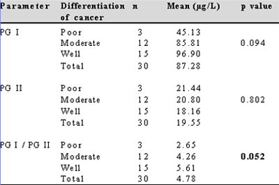
No correlation was found between PG I or PG II levels and degree of differentiation of carcinoma. However, the fall in PG I/II ratio was very nearly significant (p=0.052) (Table III).

The cut off value of serum PG I and PG I/II ratio for the diagnosis of carcinoma stomach was calculated using the ROC curve.  The cut off value of serum PG I level at 115.3 µg/L provided sensitivity and specificity of 83.3% and 66.7% respectively. The PG I/II ratio at 6.25 had a sensitivity of 87.7% and specificity of 73.3%.  When both tests, i.e. PG I and PG I/II ratio, were used in parallel i.e., only either of the two tests had to be satisfied for a positive result, the sensitivity increased to 97.0% and the negative predictive value was 91.4%.  When the two tests were applied in series, i.e. when both values had to be satisfied for the test to be considered positive, the sensitivity declined to 72.2 % and the  specificity increased to 91.9% .The likelihood ratio of a positive test (LH+) and a negative test (LH-) were1.86 and 0.06 respectively when the cut off values were applied in parallel. However, the LH+ and LH- were 8.91 and 0.30 respectively, when the cut off values were applied in series (Table IV).

Fig. 1 : PG I levels in various groups


Fig. 2 : PG I/II ratio in various groups


Fig. 3 : PG II levels in various groups

Table II: Comparison of serum PG I, PG II and PG I/II values between controls and study groups

Table III: Correlation of serum PG I, PG II and PG I/II values with the degree of differentiation of carcinoma stomach
PG-pepsinogen


DISCUSSION

Serum pepsinogen has gained much attention recently as a useful diagnostic tool in screening for gastric carcinoma.(8) However, its role in disorders like H.pylori gastritis, peptic ulcer, portal hypertensive gastropathy, erosive gastritis and non-ulcer dyspepsia is uncertain.

There is wide variation in the levels of serum PG I, PG II and PG I/II in normal individuals across different population groups.  The range varies from 40-80 µg/L for PG I, 10-15 µg/L for PG II and 5-9 for PG I/II ratio in different population groups.(9,10,11) The mean levels of PG I (158 µg/L) and PG II (22 µg/L) in normal individuals in the present study were much higher than those reported in other studies.  The ratio of PG I/II (7.2) however was similar.  The reason for a higher level of PG I and II in the population studied is outside the scope of the present study, but may be linked to the chief cell mass in different population groups.

The present study demonstrated a significant fall in the levels of PG I and PG I/II ratio in patients with carcinoma stomach.  A similar decrease in PG I and PG I/II levels in carcinoma stomach has been noted in studies across literature.(4,12) Only one study from Singapore by So et al found the ratio of PG I/II alone to be decreased in carcinoma stomach with no change in the levels of PG I or PG II.(13) The absence of a fall in PG I in their study could be due to the low prevalence of atrophic gastritis and gastric carcinoma in the population studied by them. No significant association between PG II levels and carcinoma stomach was found in the present study.  This is explained by the additional duodenal secretion of PG II which compensates for its decreased supply from the gastric source.

Reliable cut off points of PG I level and PG I/II ratio for detecting carcinoma stomach can be calculated using the ROC curve.  The calculated levels in literature range from 25-70 µg/L for PG I and 3-5 for PG I/II.(12,14) In the present study the cut off level, to diagnose carcinoma stomach, of serum PG I was 115.3 µg/L and for PG I/II ratio was 6.25. When both tests, i.e. PG I and PG I/II ratio were used in parallel the sensitivity was 97.0% and the negative predictive value was 91.4%.  Using the two tests in series the sensitivity declined to 72.2%.The sensitivity and specificity of these levels in detecting carcinoma stomach are comparable to the previous studies.

The predictive values of any diagnostic test are dependent on the prevalence of the disease in the population studied.(15) Hence to obtain a more reliable way of assessing the usefulness of a test the sensitivity and specificity can be combined into one measure called the Likelihood Ratio (LR).When test results are reported as being either positive or negative two types of LRs can be described, viz, likelihood ratio for a positive test, LR (+), and the likelihood ratio of a negative test, LR (-). A low LR(-) rules out disease.

The LR(-)value was 0.06 when the PG I level and PG I/II ratio were applied in parallel, indicating that individuals without carcinoma were 16 times more likely to have a negative test than those with carcinoma, thus making it a good screening test.
                     
Various studies have attempted to elucidate the correlation between pepsinogen levels and H.pylori infection with conflicting conclusions.(16)  Studies have found serum PG II levels to be raised in H.pylori infection and have recommended the use of serum PG II levels to diagnose and to evaluate eradication of H.pylori infection.(5,17) On the contrary other studies have found an elevated serum PG I over PG II in H.pylori infection.(18) In the present study there was no significant change in PG I, PG II or the PG I/II ratio in patients with H.pylori gastritis.  A diagnosis of H.pylori gastritis was made by urease testing alone. It is known that H.pylori can cause an increased pepsinogen level during the initial phase of mucosal inflammation with a subsequent fall in its levels as the inflammation progresses to atrophy.  Since there was no histological correlation of H.pylori gastritis with pepsinogen levels in the present study, our results may not be conclusive.

There exists no conclusive evidence regarding the association between peptic ulcer and pepsinogen level.  Studies have described elevated PG I/II ratio in duodenal ulcer.  Duodenal ulcer is also shown to have a high PG I level.4 Other studies have found increased PG I level in gastric ulcer rather than duodenal ulcer.(19) In the present study, we found no significant association between peptic ulcer and serum pepsinogen levels.

Very few studies are available on the role of pepsinogen in portal gastropathy with no demonstrable relationship between them.(20) This was our observation as well. We found no correlation between serum PG I and PG II levels with erosive gastritis and NUD. Studies done earlier have found no relationship between pepsinogen level and erosive gastritis.(9) There exists no conclusive data in the literature about the relationship between NUD and PG levels.

From this study, we conclude that estimation of serum pepsinogen level is a reliable tool in screening for carcinoma stomach. Patients with abnormal values may be selected for endoscopy. There was no significant change in serum pepsinogen levels in disorders like peptic ulcer, portal hypertensive gastropathy, NUD, erosive gastritis and H.pylori gastritis; thus the role of serum pepsinogen level in the diagnosis of these disorders was not established.

The main limitations of the present study were a  relatively small sample size, lack of severity grading of gastritis leading to an inability to correlate the levels with severity of gastritis, and inadequate number of patients in subgroups of carcinoma stomach for valid comparisons to be made between different degrees of differentiation. Also, since we had no patients with early gastric carcinoma we could not assess the utility of these values in early gastric cancer.

REFERENCES

1.     Samloff IM, Varis K, Ihamaki T, Siurala M, Rotter JI. Relationship among serum pepsinogen I, serum pepsinogen II and gastric mucosal histology: a study in relatives of patients with pernicious anemia.  Gastroenterology. 1982;83:204–9.

2.     Samloff IM.  Pepsinogen, pepsins and pepsin inhibitors.  Gastroenterology. 1971;60:586–604.

3.     Fukao A, Hisamichi S, Ohsato N, Fujino N, Endo N, Iha A. Correlation between the prevalence of gastritis and gastric cancer in Japan.  Cancer Causes Control. 1993;4:17–20.

4.     Huang SC, Miki K, Furihata C, Ichinose M, Shimizu A, Oka H.  Enzyme-linked immunosorbent assays for serum pepsinogen I and II using monoclonal antibodies – with data on peptic ulcer and gastric cancer. Clin Chim Acta. 1988;175:37–50.

5.     Di Mario F, Moussa AM, Cavallaro LG, Caruana P, Merli R,Bertolini S, et al.  Clinical usefulness of serum pepsinogen II in the management of Helicobacter pylori infection.  Digestion. 2004;70:167–72.

6.     Kikuchi S, Kurosawa M, Sakiyama T, Tenjin H. Long-term effect of smoking on serum pepsinogen levels. J Epidemiol. 2002;12:351–6.

7.     Festen HP, Thijs JC, Lamers CB, Jansen JM, Pals G, Frants RR,  et al.  Effect of oral omeprazole on serum gastrin and serum pepsinogen I level.  Gastroenterology. 1984;87:1030–4.

8.     Webb PM, Hengels KJ, Moller H, Newell DG, Palli D, Elder JB, et al.  The epidemiology of low serum pepsinogen A levels and an international association with gastric cancer rates EUROGAST Study Group.  Gastroenterology. 1994;107:1335–44.

9.     Ichinose M, Miki K, Furihata C, Kageyama T, Hayashi R, Niwa H, et al.  Radioimmunoassay of serum group I and group II pepsinogens in normal controls and patients with various disorders.  Clin Chim Acta. 1982;126:183–91.

10.   Wu MS, Lin JT, Wang JT, Huang SC, Wang CY, Wang TH.  Serum levels of pepsinogen I in healthy volunteers and patients with gastric ulcers and gastric carcinoma in Taiwan.  J Formos Med Assoc. 1993;92:711-6.

11.   Veenendaal RA, Biemond I, Pena AS, Van Duijn W, Kreuning J, Lamers CB.  Influence of age and Helicobacter pylori infection on serum pepsinogen in healthy blood transfusion donors. Gut. 1992; 33:452–5.

12.   Kitahara F, Kobayashi K, Sato T, Kojima Y, Araki T, Fujino MA.  Accuracy of screening for gastric cancer using serum pepsinogen concentrations.  Gut. 1999;44:693–7.

13.   So JB, Yeoh KG, Moochala S, Chachlani N, Ho J, Wong WK, et al.  Serum pepsinogen levels in gastric cancer patients and their relationship with Helicobacter pylori infection: a prospective study.  Gastric cancer. 2002;5:228–32.

14.   Aoki K, Misumi J, Kimura T, Zhao W, Xie T.  Evaluation of cut off levels for screening of gastric cancer using serum pepsinogens and distributions of levels of serum pepsinogen I, II and of PG I/PG II ratios in a gastric cancer case-control study.  J Epidemiol. 1997;7:143–51.

15.   Akobeng KA. Understanding diagnostic tests 2: likelihood ratios, pre-and post-test probabilities and their use in clinical practice. Acta Paediatrica. 2007;96:487–91.

16.   The EUROGAST Study Group.  An international association between Helicobacter pylori infection and gastric cancer.  Lancet. 1993;341:1359–62.

17.   Bodger K, Wyatt JI, Heatley RV.  Variation in serum pepsinogens with severity and topography of Helicobacter pylori – associated chronic gastritis in dyspeptic patients’ referred for endoscopy. Helicobacter 2001;6:216–24

18.   Mossi S, Meyer-Wyss B, Renner EL, Merki HS, Gamboni G.  Influence of Helicobacter pylori, sex and age on serum gastritis and pepsinogen concentrations in subjects without symptoms and patients with duodenal ulcers.  Gut. 1993;34:752–6.

19.   Wu MS, Wang HP, Wang JT, Wang TH, Lin JT.  Serum pepsinogen I and pepsinogen II and the ratio of pepsinogen I/II in peptic ulcer diseases: with special emphasis on the influence of the location of the ulcer crater.  J Gastroenterol Hepatol. 1995;10:401–4.

20.  Lin WJ, Lee FY, Lin HC, Tsai YT, Lee SD, Lai KH, et al.  Snake skin pattern gastropathy in cirrhotic patients.  Gastroenterol Hepatol. 1991;6:145–9.